Figures & data
Figure 1. Setup of the dodo specimen within the Zeiss Versa 520 X-ray CT scanner
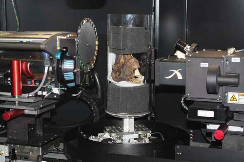
Figure 2. Ortho slices through the Oxford Dodo, Raphus cucullatus (Linnaeus, 1758) (OUMNH.ZC.11605), derived by X-ray CT scanning. The bright values indicate bone and the grey values the skin preserved on the right-hand side of the skull. (a) Sagittal section through the entire specimen; (b) Magnified view of sagittal section to show the structure of the trabecular bone; (c) Transverse section aligned through the eye region of the specimen. The sclerotic ring is visible within the skin in the top right of the image; (d) Coronal section through the posterior of the skull showing the cranial cavity; (e) Coronal section of the anterior part of the skull showing the positioning of the sclerotic ring
Figure 3. Ortho slices through the Oxford Dodo derived by X-ray CT scanning together with rendered 3D model (inset) showing position of coronal section. (a) Coronal section through the rostrum showing calcified tissue within the skin. Magnified views of this mineralised soft tissue are shown in (b) coronal section and (c) transverse section. Calcified tissue is also seen in an equivalent position in the detached skin of the left-hand side of the skull
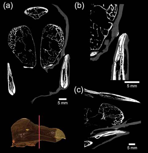
Figure 4. Rendered 3D views of the Oxford Dodo using the X-ray CT data in Drishti. Clockwise from top left: right lateral with skin; right lateral; coronal section through cranial cavity; sagittal section viewed obliquely
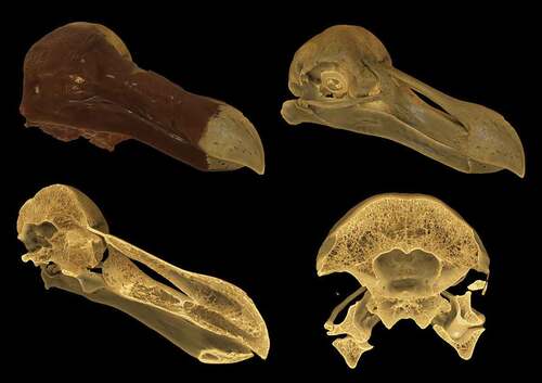
Figure 5. A colour 3D print of the Oxford Dodo skull created by generating a mesh from the X-ray CT data, with the skin of the right-hand side of the skull digitally removed prior to printing
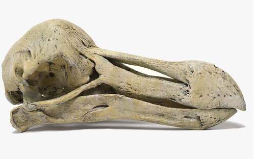
Figure 6. Ortho CT slices and rendered 3D models of the Oxford Dodo showing metal particles, characterised by star artefacts in the ortho slices, embedded within the bone of the skull, including deep within the trabecular bone. The metal particles are concentrated within the bone of the posterior left of the skull. (a) Coronal section through the cranial cavity, viewed from the posterior, with metal particles showing as bright spots; (b) Coronal section farther caudad than (a) showing a number of bright spots highlighted and enlarged to show the star artefacts and their position embedded deeply within trabecular bone; (c) 3D rendered images with the bone transparent and lead shot highlighted in white
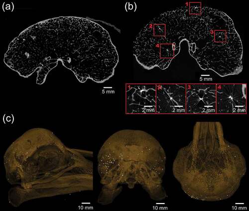
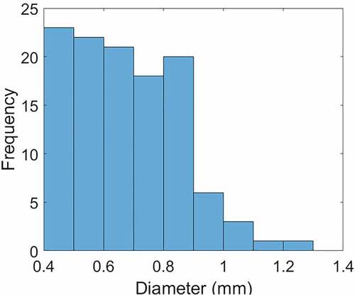
Figure 7. Histogram of the size distribution of metal particles found embedded within the specimen