
Figures & data
Figure 1. Geographical position of the locality of the Varifructus gen. nov in Liaoning, China (Drawn by X.W.). Reproduced from Han et al. (Citation2017) with permission and courtesy of Acta Geologica Sinica (English edition). (a) Fossil locality (black dot) in northeastern China. (b) Detailed position of fossil locality Dawangzhangzi (black square) in a suburb of Lingyuan, Liaoning
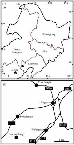
Figure 2. Distal portion of the plant of Varifructus gen. nov, Ma007. Note the nodes (1, 2), a flower bud (3), and fruits of varying morphologies (4 − 9). Bar = 2 cm
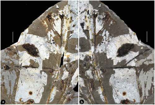
Figure 3. Branching of Varifructus gen. nov. (a) Detailed view of Node 1 in ), which exhibits opposite axillary branching. Note the two subtending bisected leaves (black arrows) and axillary branches (white arrows). Bar = 5 mm. (b) Detailed view of Node 2 in ), which shows axillary branching. Note the subtending leaf (black arrow) and axillary branch (white arrow), on the counterpart. Bar = 2.5 mm
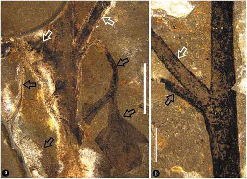
Figure 4. Fruit pair and flower bud of Varifructus gen. nov
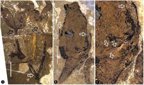
Figure 5. Fruit pair and its details
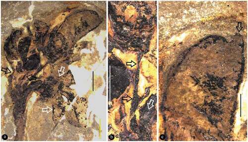
Figure 6. Fruit pair and its details
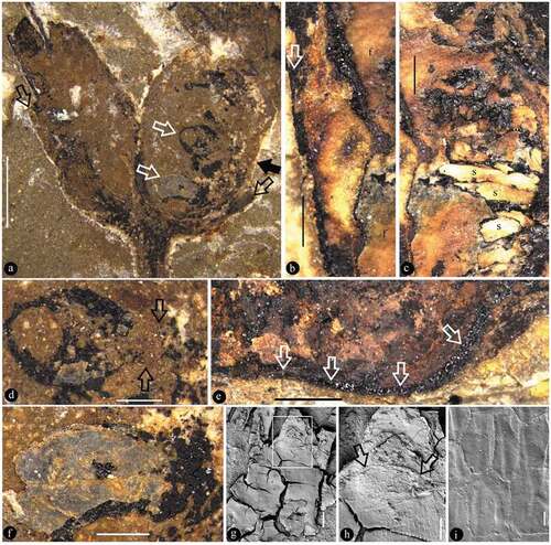
Figure 7. Fruit marked as 5 in Figure 2b (on the counterpart) and its details
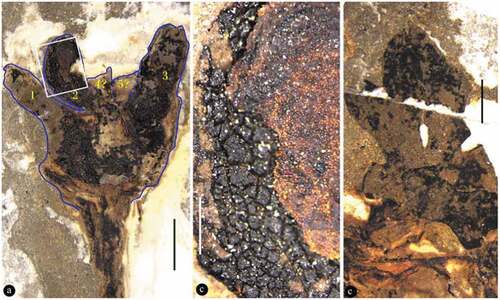
Figure 8. Reconstruction of Varifructus gen. nov
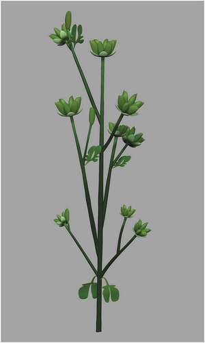
Table 1. Comparison between Varifructus gen. nov and other angiosperms from the Yixian Formation