Figures & data
Figure 1. Location and geological setting of the studied area. A, map showing the location of the research area (red star) in the Yangtze Platform of South China. B, lithofacies reconstruction of South China during the Cambrian Miaolingian (after Feng et al. Citation2004; Zhang et al. Citation2008) with the studied area indicated by a red star. C, geological map of the Balang area and location of the Wuliu-Zengjiayan, Miaobanpo sections and the Jinyinshan quarry (modified from Zhao et al. Citation2019). D, stratigraphical column of the Kaili Formation at the Miaobanpo section, with fossil horizons indicated (after Yang et al. Citation2016).
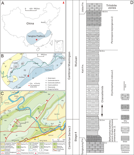
Figure 2. Terminology of chancelloriid sclerites. The lower right corner shows the schematic diagram of angle measurement between two marginal-lateral rays (modified from Bengtson and Collins Citation2015 and Yun Citation2019).
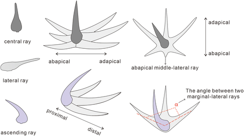
Figure 3. Archiasterella anchoriformis sp. nov. from the Kaili Biota. A, the fossil sketch drawing of B, dotted lines represent the indistinctive sclerites. B, the holotype scleritome (MBP-38); the white arrow points to an ascending ray of the sclerites. C, the sclerites of B (white arrow) and the proximal part of sclerites slightly swells. D, overall morphology, specimen no. MBP-43. E, overall morphology, specimen no. MBP-34. F, middle part of the body, specimen no. MBP-44. G, the apical tuft of MBP-43, is the countpart of D. H, J-K, the sclerites of D, E and F respectively, white arrow points to the ascending rays. I, the collapse of rays of D. Scale bars: A-B, D-F = 2 mm; C, G-H, J-K = 0.5 mm; I = 0.2 mm.
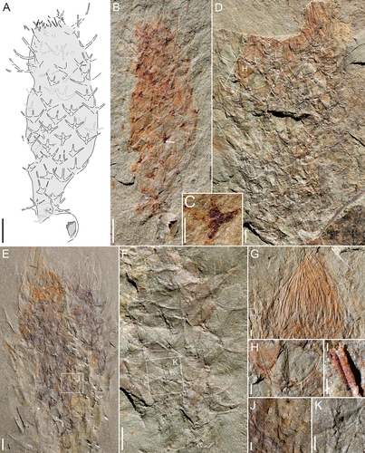
Figure 4. Archiasterella anchoriformis sp. nov. from the Kaili Biota. A, specimen no. JYS-1074. B, JYS-1074-1 of A (the lower left), 2 + 1A sclerites is present (red frame). C, JYS-1074-2 of A (the upper right). D, the specimen preserved from upper direction of A (the lower right), uncertain its type. E, detail of B (position marked by white frame). Scale bars: A = 5 mm; B-C = 2 mm; D = 1 mm; E = 0.5 mm.
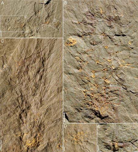
Figure 5. Chancelloria zhaoi sp. nov. from Kaili Biota. A, the fossil sketch drawing of B, dotted lines represent the uncertainty of sclerites. B, the holotype scleritome (MBP-42-1). C, apical tuft and orifice of B, the white arrow point to a stoutly composite sclerites. D, the paratype scleritome (MBP-46-3). E, 6 + 1C and 5 + 1C sclerites of C. zhaoi sp. nov., the length of ‘adr’ (adapical lateral ray) is longer than ‘abr’ (abapical lateral ray). Scale bars: A-B, D = 1 mm; C = 0.5 mm.
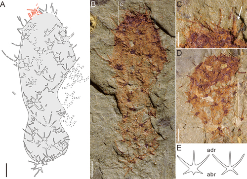
Figure 6. Chancelloria eros from the Kaili Biota. A, including four specimens of Chancelloria eros, specimen no. MBP-41. The specimen with the largest size is MBP-41-1. B, the second specimen (MBP-41-2) from the bottom of A, the white arrow indicates that the sclerite has a longer lateral ray. C, the third specimen (MBP-41-3) from the bottom of A, the white arrow stands for the possibility of stalk structure, and the red arrow indicates that the basal part of MBP-41-3 probably anchored to the top of MBP-41-2. D, the fourth specimen (MBP-41-4) from the bottom of A. E-G, 6 + 1C, 5 + 1C and 7 + 1C sclerites of the first C. eros from the bottom of A. Scale bars: A = 5 mm; B-D = 2 mm; E-G = 0.5 mm.
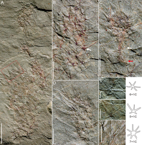
Figure 7. Allonnia from Kaili Biota. A-D, F, Allonnia erjiensis. A, the fossil sketch drawing of B, dotted lines represent the uncertainty of sclerites. B, MBP-35. C, MBP-45. D, MBP-37. E, G, Allonnia phrixothrix, specimen no. MBP-40. F, detail of D (position marked by white frame). G, integument detail of E; ‘lr’, lateral ray; ‘asr’, ascending ray. Scale bars: A-E = 2 mm; F, G = 0.1 mm.
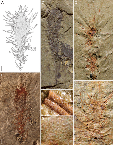
Figure 8. Allonnia sp. from Kaili Biota. A, three specimens in different direction, specimen no. MBP-47. The white box indicates the basal part of MBP-47-2 probably anchored to the top of MBP-47-3. B, apical tuft of the lower right corner of A. C, sclerites detail of the upper left corner of A. D, MBP-39. E, MBP-49; ‘lr’, lateral ray; ‘asr’, ascending ray. Scale bars: A = 5 mm; B, C = 0.2 mm; D, E = 2 mm.
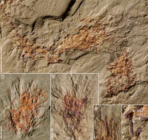
Figure 9. The reconstruction of chancelloriids in Kaili Biota (By Liu X). The labels 1 to 5 indicate Allonnia erjiensis, Chancelloria eros, C. zhaoi sp. nov., Archiasterella anchoriformis sp. nov., and Al. phrixothrix, respectively.
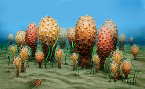
Figure 10. Distribution of the chancelloriids in South China.The stratigraphical distribution, shows two flourishing ages of genus-level chancelloriids. References for different formation: Kuanchuanpu Fm. = Fu (Citation1983), Yang et al. (Citation1983) and Steiner et al. (Citation2004); Huangshandong Fm. = Qian et al. (Citation1979) and Qian (Citation1989); Maidiping Fm. = Yin et al. (Citation1980), Qian (Citation1989), Li (Citation1999), Feng et al. (Citation2022); Zhongyicun to Dahai Mbs. of Zhujiaqing Fm. and Shiyantou Fm. = Luo et al. (Citation1982), Parkhaev and Demidenko (Citation2010), Moore et al. (Citation2014); Middle Jiulaodong Fm. = Yin et al. (Citation1980) and Qian (Citation1989); Xihaoping Fm. = Qian and Zhang (Citation1983), Duan (Citation1984), Wang and Xu (Citation1987), Qian (Citation1989), Ding et al. (Citation1990), Li et al. (Citation2004), Steiner et al. (Citation2004), Moore et al. (Citation2010), Yang et al. (Citation2015), Zhang et al. (Citation2021) ; Lower Shuijingtuo Fm. = Li et al. (Citation2004) and Yang et al. (Citation2015); Qingjiang Biota = Fu et al. (Citation2019); Chengjiang Biota = Bengtson and Hou (Citation2001), Janussen et al. (Citation2002), Hou et al. (Citation2014), Yun et al. (Citation2018), Cong et al. (Citation2018), Sun et al. (Citation2020), Yun et al. (Citation2022); Upper Jiulaodong Fm. = Yin et al. (Citation1980); Middle Guojiaba Fm. = Yun et al. (Citation2021) ; Lower Mingxinsi Fm. = Qian (Citation1989); Guanshan Biota = Hu et al. (Citation2010, Citation2013), Chen et al. (Citation2019), Zhao et al. (Citation2018); Mufushan Fm. = Zhang (Citation1995); Balang Fm. = Liu and Lei (Citation2013); Kaili Biota = Zhao et al. (Citation1999, Citation2005, Citation2011), Harvey et al. (Citation2012), this study; Upper Damao Gp. = Jiang and Huang (Citation1986), Damao Group is distributed in Hainan and likely near to the Yangtze Block in the Cambrian; Huaqiao Fm. = Zhu et al. (Citation2003).
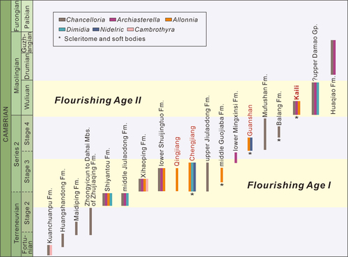
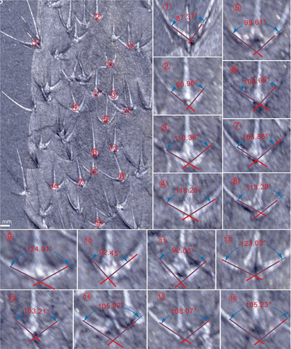
Figure A1. Archiasterella anchoriformis of Kaili Biota (MBP-38). The average angle of this specimen is 119.9°.
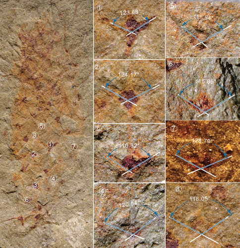
Figure A2. Archiasterella anchoriformis of Kaili Biota (MBP-43). The average angle of this specimen is 127.7°.
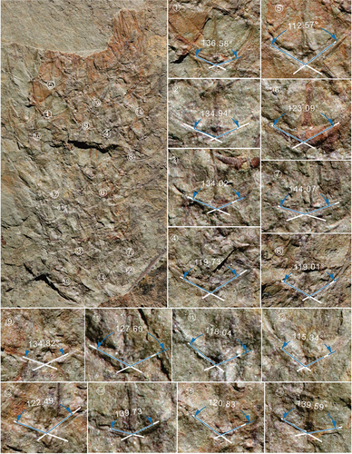
Figure A3. Archiasterella anchoriformis of Kaili Biota (JYS-1074-1). The average angle of this specimen is 130.8°.
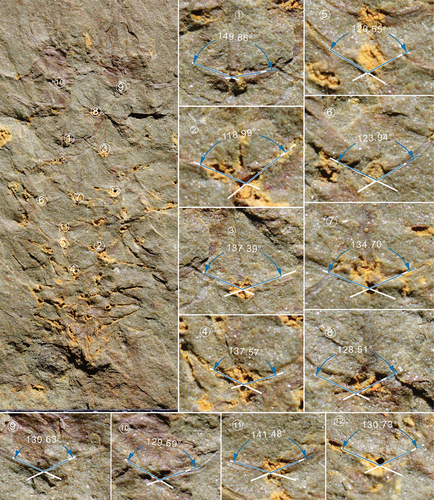
Figure A4. Ar. coriacea of Burgess Shale (based on Bengtson and Collins, 2015). The average angle of this specimen is 105.1°.