Figures & data
Figure 1. Geographical and geological maps. A, map of Scandinavia with location of allochthonous sections along the Caledonian front and distribution of surface and subsurface Lower Palaeozoic rocks (modified from Nielsen and Schovsbo Citation2011; Ebbestad et al. Citation2022). B, map of Scania, southern Sweden with Cambrian outcrop areas and localities discussed herein. C, map of the Brantevik–Gislövshammar area with location of the sites sampled for this study and the type section of the Gislöv Formation.
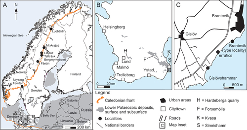
Figure 2. Stratigraphy and lithology of the Gislövshammar section, with previous trilobite zonation (left) and revised trilobite zonation (right) (modified from Bergström and Ahlberg Citation1981, fig. 8; Cederström et al. Citation2022, fig. 7).
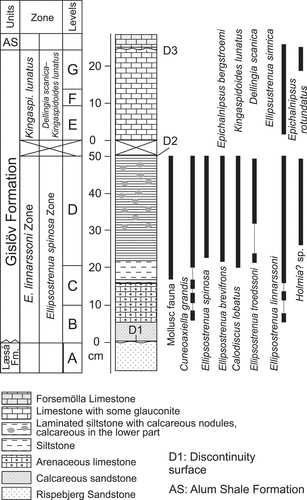
Figure 3. A–F, size frequency distribution of helcionelloid molluscs and Calodiscus sclerites from the Gislövshammar section. A, Helcionella antiqua. B, Stenotheca norvegica. C, Davidonia puppis. D, Davidonia cf. rostrata. E, Calodiscus lobatus (cephala). F, Calodiscus lobatus (pygidia). G, H, rarefaction diversity calculated from the Brantevik, Hardeberga, Gislövshammar and Mjøsa sections with trilobites only (G) and trilobites and helcionelloids combined (H). The x axis has been truncated to save space as the number of specimens exceeds 2200.
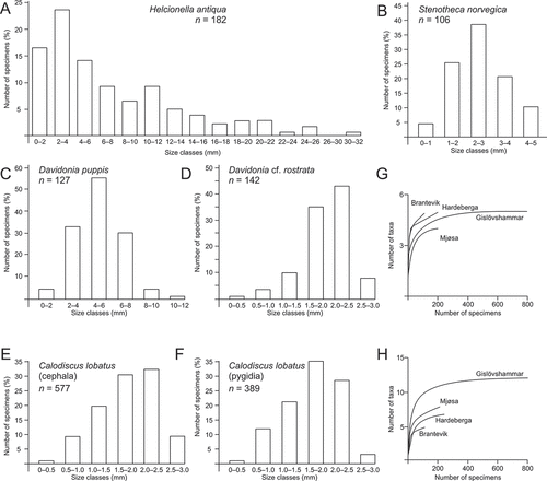
Figure 4. Quantitative visualisation of associations of taxa in Ellipsostrenua spinosa Zone in Norway and Sweden (paired group algorithm and the Dice similarity index). A, cluster analysis of trilobites only, with upper and lower beds of the Mjøsa and Mount Assjatj sections combined. B, cluster analysis of trilobites only, with upper and lower beds of the Mjøsa and Mount Assjatj sections separated. C, cluster analysis of trilobites and helcionelloids, with upper and lower beds of the Mjøsa and Mount Assjatj sections combined. D, cluster analysis of trilobites and helcionelloids, with upper and lower beds of the Mjøsa and Mount Assjatj sections separated.
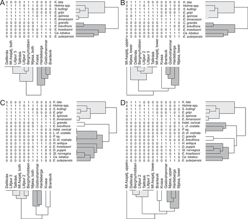
Table 1. Distribution and number of specimens of helcionelloid molluscs and associated trilobites at six Scandinavian localities (see ), and associated diversity calculations for each locality. Data on trilobites from Cederström et al. (Citation2012, Citation2022).
Figure 5. Tentative correlation of the Ellipsostrenua spinosa Zone in Sweden and Norway. The range of helcionelloid molluscs indicates a strong correlation between the Gislövshammar and Mjøsa sections. The Mount Assjatj section is omitted for reasons of space as the interval is several metres thick compared to the centimetre scale for the other sections. See. for location of sections. Data added from Cederström et al. (Citation2012, Citation2022) and Høyberget et al. (Citation2015)
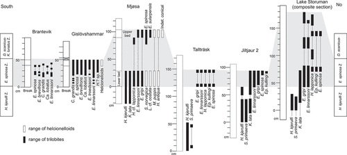
Figure 6. ‘Pelagiella’ sp. A, external mould showing ornamentation (PMU 37517). B, C, dorsal and dorsal oblique view of internal mould (PMU 37518/1). D, internal mould (PMU 37524). E, F, lateral oblique and lateral views of internal mould (PMU 37520). From the Ellipsostrenua spinosa Zone at Gislövshammar, Scania, southern Sweden. Scale bars = 1 mm.
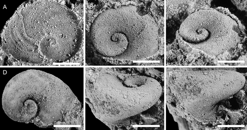
Figure 7. Helcionella antiqua (Kiær, Citation1917), with specimens arranged in an ontogenetic series. A, F, dorsal and left lateral view (PMU 37554). B, G, dorsal and posterior views (PMU 37555). C, H, dorsal and left lateral views (PMU 37556). D, E, I, dorsal,left lateral and posterior views (PMU 37557). J, K, L, dorsal, posterior oblique and detail of apex views (PMU 37558). From the Ellipsostrenua spinosa Zone at Gislövshammar, Scania, southern Sweden. Scale bar in D is 1 mm, and is common to all images.
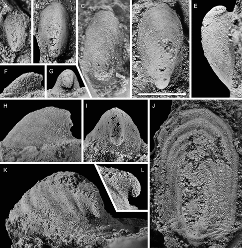
Figure 8. Helcionella antiqua (Kiær, Citation1917), with specimens arranged in an ontogenetic series. A–D, dorsal, left lateral, anterior and anterio-dorsal oblique views (PMU 37559). E, dorsal view (PMU 37561). F, dorsal view (PMU 37562/1). G–I, dorsal, posterior and detail views (PMU 37560). From the Ellipsostrenua spinosa Zone at Gislövshammar, Scania, southern Sweden. Scale bar in F = 1 mm and is common to all images.
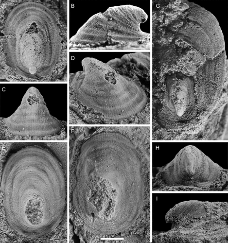
Figure 9. Large specimens of Helcionella antiqua (Kiær, Citation1917). A–C, dorsal, posterior and right lateral views (PMU 37563). D, G, dorsal and right lateral views of large specimen (PMU 37564/1) next to a medium sized specimen (PMU 37564/2). E, F anterior and dorsal views (PMU 37565). From the Ellipsostrenua spinosa Zone at Gislövshammar, Scania, southern Sweden. Scale bar in B = 10 mm and is common to all images.
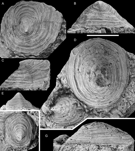
Figure 10. Specimens from a loose block collected between Brantevik and Gislövshammar, Scania, southern Sweden. A–E, Helcionella antiqua (Kiær, Citation1917). A–C, dorsal, posterior oblique and right lateral views (PMU 38127). D, E, right lateral and dorsal views (PMU 38128). F, Davidonia puppis (Høyberget et al., Citation2015). Left lateral view (PMU 38133). From the Ellipsostrenua spinosa Zone. Scale bars = 5 mm.
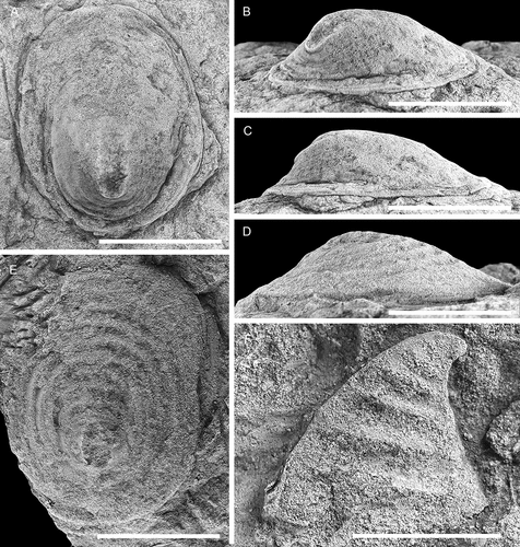
Figure 11. Comparison between Scenella barrandei (Linnarsson, Citation1879) and Helcionella antiqua (Kiær, Citation1917), showing dorsal, anterior, and lateral views and detail of shell ornamentation. A, B, E, F, Scenella barrandei, holotype (SGU 4532a), from the middle Cambrian Exsulans Limestone bed (Paradoxides paradoxisimus Zone) of the Alum Shale Formation at Kiviks-Esperöd in Scania, southern Sweden. C, D, G, H, Helcionella antiqua (PMU 37777) from the Ellipsostrenua spinosa Zone at Hardeberga, Scania, southern Sweden. Scale bars for A–E, H = 5 mm, scale bars for F, G = 1 mm.
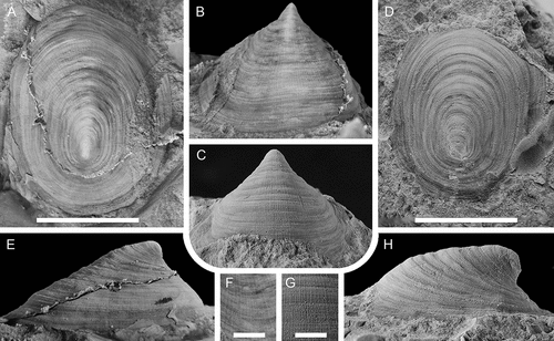
Figure 12. Davidonia puppis (Høyberget et al., Citation2015), with specimens arranged in an ontogenetic series. A, D, right lateral and anterior views (PMU 37862). B, right lateral view (PMU 37863). The white arrows point to the thin outer shell. C, right lateral view (PMU 37864). E, dorsal view (PMU 37865/2). F, G, right lateral and dorsal views (PMU 37866). H, right lateral view (PMU 37867). I, right lateral view (PMU 37868). From the Ellipsostrenua spinosa Zone at Gislövshammar, Scania, southern Sweden. Scale bars = 1 mm.
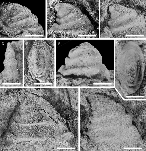
Figure 13. Davidonia puppis (Høyberget et al., Citation2015), with specimens arranged in an ontogenetic series. A, B, left lateral view and detail of posterior knobs (PMU 37869/1). Arrows in A point to thin outer shell layer; arrow in B points to shell material filling the space between knobs. C, left lateral view (PMU 37870). D, left lateral view (PMU 37871). E, right lateral view (PMU 37872). F, right lateral view (PMU 37873). G, left lateral view (PMU 37874). From the Ellipsostrenua spinosa Zone at Gislövshammar, Scania, southern Sweden. Scale bars = 1 mm.
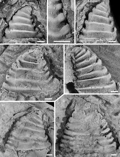
Figure 14. Davidonia puppis (Høyberget et al., Citation2015), showing details of shell morphology, shell mineralogy and ornamentation. A, B, right lateral and dorsal views (PMU 37875). C, dorsal view (PMU 37877). D, detail of ornamentation (external mould) (PMU 37876). E, F, anterior and posterior views (PMU 37878). G, H, silicon cast showing partly preserved shell ornamentation (PMU 37879). I, J, right lateral view of large internal mould with partially preserved shell layers (PMU 37880). K–M, dorsal, right lateral and posterior views of specimen with inner shell layer preserved (PMU 37881). From the Ellipsostrenua spinosa Zone at Gislövshammar, Scania, southern Sweden. Scale bars = 1 mm.
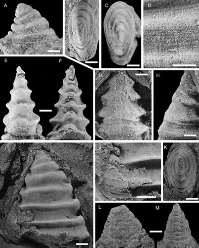
Figure 15. Davidonia cf. rostrata (Zhou & Xiao, Citation1984), with specimens arranged in an ontogenetic series. A, right lateral view (PMU 38253). B, right lateral view (PMU 37983). C, left lateral view (PMU 37984). D, right lateral view (PMU 38288). E, left lateral view (PMU 37987). F, G, right lateral and dorsal oblique views (PMU 37988/1). H, I, right lateral and anterior oblique views (PMU 37989). Arrow points to outer shell layer. J–L, left lateral, posterior and dorsal view of specimen with inner shell layer preserved (PMU 37992). M, left lateral view (PMU 37990). N, right lateral view (PMU 37991). From the Ellipsostrenua spinosa Zone at Gislövshammar, Scania, southern Sweden. Scale bars = 1 mm.
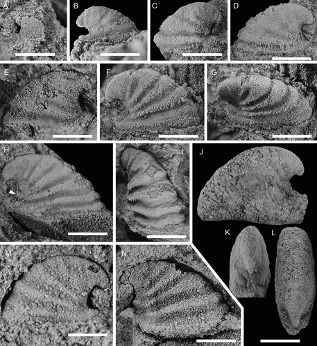
Figure 16. Davidonia cf. rostrata (Zhou & Xiao, Citation1984), showing details of morphology and shell preservation. A, B, left lateral view and detail of shell layers (PMU 37993/2). Arrows points to outer shell layer. C, left lateral view (PMU 37994/1). D, E, left lateral view and detail of shell layer (PMU 37993/1). F, right lateral view. Arrow points to outer shell layer (PMU 37995). G, left lateral view of specimen with partly recrystallised shell (PMU 38252). Arrow points to outer shell layer, obscuring the ribs on the internal mould. H, left lateral view of specimen showing two layers of recrystallised shell (PMU 38194/1). I, right lateral view of specimen showing inner shell structure of oblique bands (PMU 38289). From the Ellipsostrenua spinosa Zone at Gislövshammar, Scania, southern Sweden. Scale bars for A, C, D, F–I = 1mm, scale bars for B, E = 0.5 mm
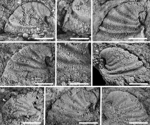
Figure 17. Latouchella cf. costata Cobbold, Citation1921, with specimens arranged in an ontogenetic series. A, left lateral view (PMU 38089). B, left lateral view (PMU 38090/1). C, right lateral view (PMU 38091). D–F, left lateral, posterior oblique and dorsal views (PMU 38092). G posterior view (PMU 38093). H, left lateral view (PMU 38094). From the Ellipsostrenua spinosa Zone at Gislövshammar, Scania, southern Sweden. Scale bars = 1 mm.
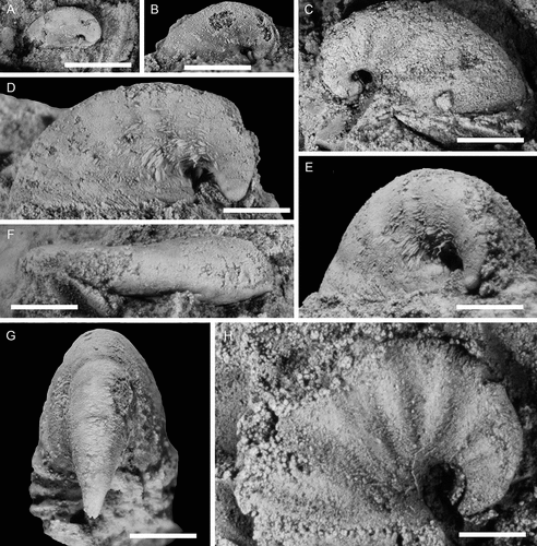
Figure 18. Stenotheca norvegica (Resser, Citation1938), with specimens arranged in an ontogenetic series. A, right lateral view (PMU 37778). B, right lateral view (PMU 37779). C, right lateral view (PMU 37780). D, right lateral view (PMU 37781). E, left lateral view (PMU 37782). F, right lateral view (PMU 37783/1). G–;J, anterior oblique, right lateral, dorsal, and dorsal oblique views (PMU 37784). K, silicon cast of specimen showing ornamentation, left lateral view (PMU 37785). L, left lateral view, internal mould (PMU 37786). From the Ellipsostrenua spinosa Zone at Gislövshammar, Scania, southern Sweden. Scale bars = 1 mm.
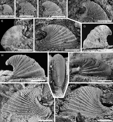
Figure 19. Stenotheca norvegica (Resser, Citation1938), with specimens arranged in an ontogenetic series. A, external mould, left lateral view (PMU 37787). B, left lateral view (PMU 37778), C, D, right lateral and anterior views (PMU 37789/1). E, external mould, ventral view, showing key-hole morphology (PMU 37790). F, silicon cast of large specimen (PMU 37791). G, silicon cast showing detail of shell ornamentation (PMU 37792/1). From the Ellipsostrenua spinosa Zone at Gislövshammar, Scania, southern Sweden. Scale bars = 1 mm

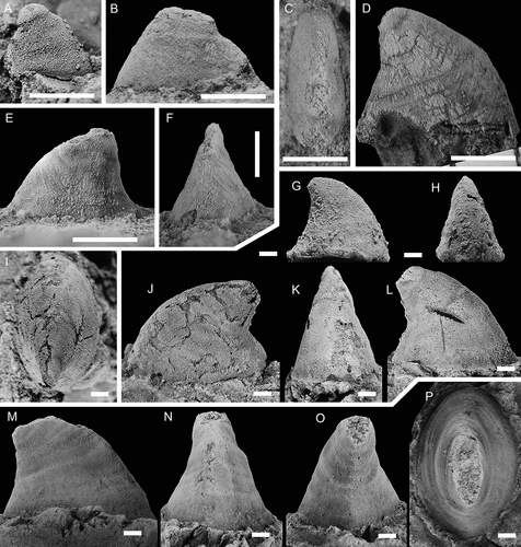
Figure 20. Conical Helcionelloid gen. and sp. indet., with specimens arranged in an ontogenetic series. A, right lateral view (PMU 37518/2). B, C, left lateral and dorsal views (PMU 38107). D, right lateral view (PMU 38108). E, F, left lateral and anterior views (PMU 38109). G, H, right lateral and anterior views (PMU 38110). I–L, dorsal, left, anterior and right lateral and views (PMU 38112). M–P, right lateral, anterior, posterior and dorsal views (PMU 38113). From the Ellipsostrenua spinosa Zone at Gislövshammar, Scania, southern Sweden. Scale bars in A–H = 1 mm, scale bars in I–P = 5 mm.