
Figures & data
Table 1. Preserved skull elements, damage and taphonomic distortion in recently recovered Genyornis newtoni skull material from Lake Callabonna. Note that all of these specimens are also associated with post-cranial material not described here.
Figure 1. The near-complete, articulated skull of Genyornis newtoni (SAMA P59516): A. Left lateral view; B. Left lateral view outlined with major parts distinguished; C. Right lateral view; D. Right lateral view outlined with major parts distinguished; E. Dorsal view; F. Caudal view; G. Ventral view; H. Rostral view. Annotations: cb., ceratobranchial; cr., cranium; ma., mandible; or., orbit; pt., pterygoid; ro., rostrum; qu., quadrate; ve., articulated vertebrae. Scale bars: 50 mm, E. to G. all to same scale.
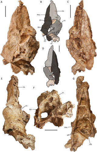
Figure 2. Genyornis newtoni caudal orbit, NMV P256893, rostral view showing the caudal orbit and quadrate articulation with the skull and mandible: A. Image; B. Annotated outline. Annotations: ap.zyg.oss., aponeurosis zygomatica ossificans; cr.or., crista supraorbitalis; f.pseu., fossa pseudotemporalis; mand., mandible; m.lev., origin for m. levator palpebrae dorsalis; n.for., foramen neurovasculare; quad., quadrate. Scale bar: 10 mm. Dark grey shading indicates regions where damage precludes morphological assessment, and light grey indicates foramina and fossae. Dotted lines provide approximate region corresponding to labelled area, and do not indicate exact boundaries.
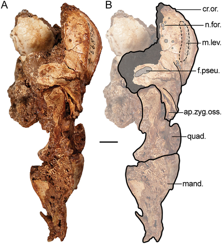
Figure 3. Lateral view of the left side of the cranium of Genyornis newtoni: A. SAMA P59516 with shaded indication of focus region; B. SAMA P59516 image and annotated outline, digitally removed from the image of the entire skull; C. NMV P256893 image (includes part of the mandible and quadrate ventrally) and annotated outline; D. Left rostrolateral view of Anhima cornuta specimen NMV B12574, box denotes region of focus in E., Quadrate disarticulated; E. Rostrolateral view of the left lateral cranium of Anhima cornuta specimen NMV B12574. Annotations: an.tymp., annulus tympanicus; ap.dep.m., an aponeurotic site of origin of m. depressor mandibulae; ap.zyg.oss., aponeurosis zygomatica ossificans; cr.zyg., crista zygomatica; cr.nu.trans., crista nuchalis transversa; cr.or., crista supraorbitalis; cr.art., crista aponeurosis articularis; f.pseu., fossa pseudotemporalis; imp.dm., impressio m. depressor mandibulae; imp.sup., impressio m. AME superficialis; jug., jugal arch; mand., mandible; m.a.e., osseous meatus acusticus externus; pr.par., processus paroccipitalis; pr.post., processus postorbitalis; pr.sup., processus suprameaticus; quad., quadrate; t.pseu., tubercle for m. pseudotemporalis (specifically aponeurosis pseudotemporalis superficialis). Scale bars: A. 50 mm, B., C. 20 mm, D., E. 10 mm. Dark grey shading indicates regions where damage precludes confident morphological assessment, and light grey indicates foramina and fossae. Dotted lines provide approximate regions corresponding to labelled areas, and do not represent accurate boundaries.
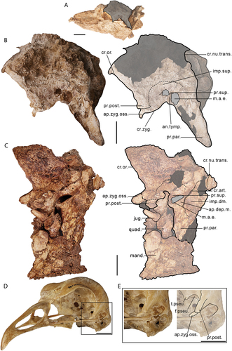
Figure 4. Soft-tissue attachment sites on the cranium of a dromornithid, illustrated using a digitally modified representation of Ilbandornis woodburnei (QMV 2000:gfv:20) due to the relative lack of deformation in this skull and the conservative nature of dromornithid skull morphology: A. Rostral aspect; B. Caudal aspect; C. Ventral aspect; D. Right lateral aspect; E. Oblique (rostrolateral) aspect. Abbreviations: ap.art., origin site of aponeurosis articularis; ap.dep.m., an aponeurotic site of origin of m. depressor mandibulae; ap.med.sup., origin site of aponeurosis mediosuperficialis; ap.sup., origin site of aponeurosis superficialis; ap.zyg., origin site of aponeurosis zygomatica (muscle fibres of m. AME profundus, pars zygomaticus, and m. AME profundus, pars superficialis, originate from the medial and lateral surfaces of this aponeurosis, respectively); lig.oc.mand., attachment area corresponding to the origin of ligamentum occipitomandibulare, continuous with that of membrana postmeatica; lig.post.orb., attachment area of origin of ligamentum postorbitale; m.AME.prof.cor., origin area of m. AME profundus, pars coronoideus; m.AME.sup., origin area of m. AME superficialis; m.biv.cerv., insertion area of m. biventer cervicis; m.c.c., origin area of m. cucullaris capitis; m.comp., insertion area of m. complexus; m.dep.mand., origin area of m. depressor mandibulae; m.pro.pter.quad., origin area of m. protractor pterygoidei et quadrati; m.ps.tem.s., origin area of m. pseudotemporalis superficialis; m.rec.cap.dors., insertion areas for slips of m. rectus capitis dorsalis; m.rec.cap.lat., insertion area of m. rectus capitis lateralis; m.rec.cap.vent.lat., area for aponeurosis of insertion of m. rectus capitis ventralis, pars lateralis; m.rec.cap.vent.med., insertion area of m. rectus capitis ventralis, pars medialis; m.spl.cap., insertion area of m. splenius capitis; mem.at.oc.dors., attachment area of membrana atlantooccipitalis dorsalis; mem.at.oc.vent., attachment area of membrana atlantooccipitalis ventralis; mem.pm., attachment site of membrana postmeatica, which incorporates and is not separatable with respect to the more ventromedial ligamentum occipitomandibulare. Scale bar: 50 mm. Illustrated attachment areas on the cranium are non-extensive estimates; only the sites corresponding to selected muscles, aponeuroses, membranes and ligaments are indicated.
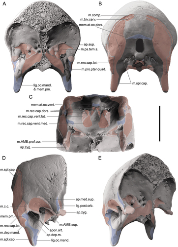
Figure 5. Caudal and ventral views of the cranium of Genyornis newtoni, digitally cropped from the images of the entire skull: A. SAMA P59516 image and annotated outline in caudal view; B. SAMA P59516 fossa parabasalis and associated foramina for nerves, focus region outlined in A.; C. Complete ventral view of SAMA P59516 with box indicating region featured in D.; D. Ventral cranium, SAMA P59516 image and annotated outline, rotated anti-clockwise from C. Annotations: can., canal tentatively identified as canalis orbitalis (see Baumel and Witmer Citation1993: annot. 95); cfp., crista fossae parabasalis; coe., ostium canalis ophthalmici externi; cond.oc., condylus occipitalis; cr.bas.lat., crista basilaris lateralis; cr.nu.trans., crista nuchalis transversus; cr.ven., crista ventralis; f.para., fossa parabasalis; fng., foramen n. glossopharyngealis (IX); fnh., foramen n. hypoglossi (XII); fnv., foramen n. vagi (X); inc.mc., incisura mediana condyli; for.mag., foramen magnum; i.para., lamina parasphenoidalis; m.rec., insertion for musculus rectus capitis dorsalis; occ., ostium canalis carotici and branch of nerve VII; pr.basi., processus basipterygoideus; pr.par., processus paroccipitalis; prom.ex., prominentia exoccipitalis; r.para., rostrum parasphenoidalis (basis rostri parasphenoidalis); s.int., septum interorbitale; tu.au., tuba auditiva communis; tubc.bas., tuberculum basilare. Scale bars: A., D. 20 mm, B. 10 mm, C. 40 mm. Dark grey shading indicates regions where damage precludes morphological assessment, and light grey indicates foramen and fossae. Dotted lines in D. indicate the alignment and distortion of the sagittal plane of each region.
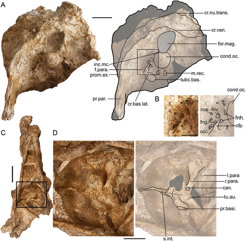
Figure 6. Genyornis newtoni right quadrate, NMV P256893: A. Lateral view, image and annotated outline; B. Medial view, image and annotated outline; C. Dorsal view; D. Ventral view; E. Rostral view; F. Caudal view. Annotations: cap.s-o., capitulum squamoso-oticum; cond.l., condylus mandibularis lateralis; cond.m., condylus mandibularis medialis; con.pt., condylus pterygoideus including facies articularis pterygoidei; cr.med., crista medialis; cr.tymp., crista tympanica; d.caud., depressio caudomedialis; d.rost., depressio rostromedialis; d.sup., depressio supracondylaris; for.rost., foramen pneumaticum rostromediale; f.basi., fossa basiorbitalis; fov.q., fovea quadratojugalis; musc., tubercles for insertion of m. protractor pterygoidei et quadrati; o.cap., otic part of capitulum squamoso-oticum; p.oti., pars otica; p.sub., prominentia submeatica; s.cap., squamosal part of capitulum squamoso-oticum; t.sub., tuberculum subcapitulare; v.inco., vallecular intercotylaris. Scale bars: 10 mm. Dark grey shading indicates regions where damage precludes morphological assessment, and light grey indicates the foramen pneumaticum rostromediale.
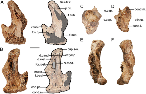
Figure 7. Genyornis newtoni rostrum, lateral views: A. SAMA P59521 left lateral view image and annotated outline; B. SAMA P59521 right lateral view; C. SAMA P59517 left and right lateral views. Annotations: ang., angulus tomialis; ca., casque; cr.tom., crista tomialis; fov., foveae corpusculorum nervosorum; lac., lacrimal ; margo.lat., margo lateralis palatini; nas., apertura nasi ossea; pala., palatinum; p.o.l., processus orbitalis of lacrimale; pr.jug., processus jugalis of the maxillare; pr.max., processus maxillopalatinus of the maxillare; pr.n.m., processus nasalis of the maxillare; sul.para., sulcus paratomialis. Scale bars: 20 mm. Dark grey shading indicates regions where damage precludes morphological assessment, and light grey indicates fenestrae, foveae, and aperturae.
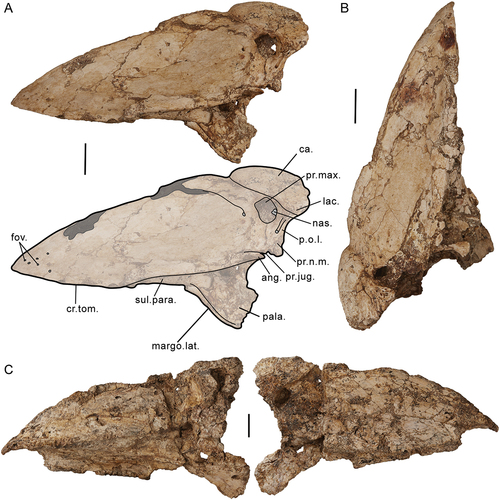
Figure 8. Dromornis planei rostrum, specimen NTM P932–2, shows distortion and cavitation on the surface of the bone: A. Right side; B. Left side; C. Close-up image of the distortion on the rostrodorsal-most part of the left side of the rostrum. Annotations: ar.jug., arcus jugalis; cr.tom., crista tomialis; fen., fenestra; lac., lacrimal; p.o.l., processus orbitalis of lacrimal. Scale bars: A., B. 50 mm and C. 20 mm.
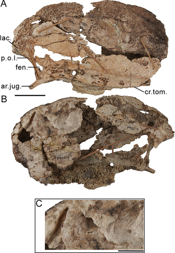
Figure 9. Genyornis newtoni rostrum, specimen SAMA P59521, and comparison of palatine and jugal arch morphology: A. Ventral/Palatal view image and annotated outline; B. Dorsal view image and annotated outline; C. Caudal view image and annotated outline; D. Hypothetical reconstruction of the palatines of Genyornis newtoni (pale orange), pars lateralis palatini (dark orange), and the jugal arch (brown) from specimen SAMA P59521; E. the galliform condition, Ortalis canicollis (see Zusi and Livezey Citation2006: fig. 7A); F. the anseriform condition, Anser albifrons (see Zelenkov and Stidham Citation2018: fig. 2A). Annotations: ang., angulus tomialis; bulb., bulbous regions largely associated with each palatine including complete synostosis with the processus maxillopalatinus of the maxillare, the processus rostralis of the palatines, and the processus palatinus of the praemaxillary; ca., casque; cho.na., choana nasalis ossea; cr., craniorostral hinge; cr.tom., crista tomialis; cul., culmen; fen.pal., fenestrae palatina; fos.cho., fossa choanalis palatini; fov., foveae corpusculorum nervosorum; lac. – lacrimal; iam.d. – lamella dorsalis, pars choanalis palatini; margo.lat., margo lateralis palatini; os.pal., ossa palatina; pal., palatum osseum; pons.mj. – pons maxillaro-jugalis; pr.cho. – processus choanalis palatini; pr.jug., processus jugalis of the maxillare; pr.max., processus maxillopalatinus; sep.na., septum nasale osseum; sn.pl., supranarial plate (of processus maxillopalatinus); sul.para., sulcus paratomialis and associated depression; sut.Vm., sutura vomeromaxillaris; vom., vomer. Scale bars: 20 mm, D.-F. not to scale. Dark grey shading indicates regions where damage precludes morphological assessment, light grey indicates sulci, fenestrae, foveae, and aperturae, dotted lines follow the rostral continuation of the sulcus paratomialis, and the white arrows in B and C indicate the lateral articular ‘condyles’ (internal processes of the nasolacrimals, sensu Murray and Vickers-Rich Citation2004).
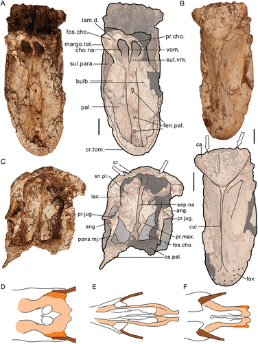
Figure 10. Genyornis newtoni mandible morphology: A. Caudal view of skull part NMV P256893, partially preserving ramus mandibulae, pars caudalis ventrally, which retains the medial process of the mandible; B. Image and annotated outline of SAMA P59516 in right lateral view, digitally removed from the image of the entire skull; C. Ventral view of the symphysial region of mandible specimen SAMA P59516; D. Caudal view of symphysial part of mandible specimen NMV P256893; E. Dorsal view of symphysial part of mandible specimen NMV P256893; F. Proposed arrangement of insertions of the adductor muscle complex on the lateral side of the mandible for dromornithids (drawn from specimen NTM P2774–2 of Ilbandornis sp. due to the limited distortion and conservative nature of dromornithid skull morphology) compared with re-drawn examples of extant galloanserans, Anseres – Anas platyrhynchos (Davids Citation1952a: fig. 3b), Anhimidae – Anhima cornuta (NMV B12574), Megapodiidae – Aepypodius arfakianus (Weber Citation1996: fig. 2a) and Phasianidae – Gallus gallus (Fujioka Citation1963: fig. 7). Annotations: c.lat., cotyla lateralis.; cr.par., crista paracoronoidea rostralis and crista paracoronoidea caudalis.; ADM, angulus dorsalis mandibulae; AVM, angulus ventralis mandibulae; exc., excavation associated with the region of recessus conicalis in anatids; f.neur., foramen neurovascularis.; m.AMEPc, m. AME profundus, pars coronoideus; m.AMEs, m. AME superficialis; m.AMEPs, m. AME profundus, pars superficialis; m.AMEPz, m. AME profundus, pars zygomaticus; m.AMP, m. adductor mandibulae posterior; m.ps., abraded prominence and approximate area for insertion of m. pseudotemporalis profundus; pr.cor., processus coronoideus (approximate position indicated, not visible as it is obscured by associated cranium); pr.lat.m., processus lateralis mandibulae; pr.med.m., processus medialis mandibulae; pr.ret., processus retroarticularis; sym., pars symphysialis, rostrum mandibulae; tom., crista tomialis; Scale bars: A., C.-E. 10 mm, B. 20 mm, F. not to scale. Dark grey shading indicates regions where damage precludes morphological assessment, and light grey indicates fenestra.
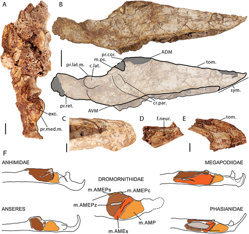
Figure 11. The hyoid elements of Genyornis newtoni specimen SAMA P59516: A. Complete specimen SAMA P59516 in ventral view; B. paraglossum, arrows for orientation (dorsal, ventral); C. Right lateral view of the basihyal and both left and right ceratobranchials in situ, image rotated 90 degrees clockwise from A.; D. Annotated outline of hyoid elements, arrow for orientation of basihyal only (clockwise: dorsal, rostral, ventral, caudal). Annotations: a.cb., damaged region just rostral of the articulatio ceratobasihyalis; a.pb., surface corresponding to articulatio paraglosso-basihyalis; bh., basihyal; cb., ceratobranchials; cor., cornua (processus paraglossus caudalis); for., central foramen. Scale bars: A. 40 mm, B. 10 mm, C., D. 20 mm.
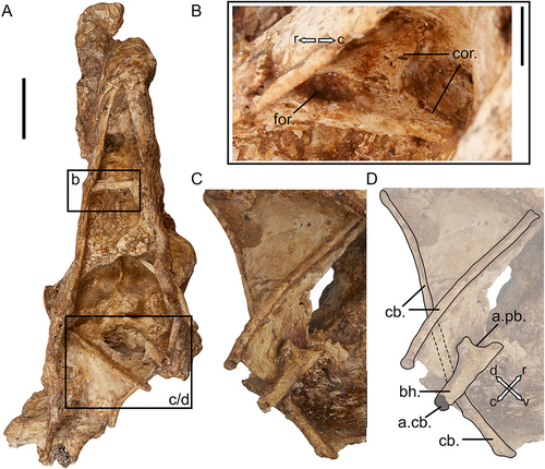
Figure 12. Galloanseran consensus trees derived from skull characters to assess the relationships of dromornithids: A. Parsimony strict consensus tree cladogram of 2 MPTs (length = 786, corresponding phylograms are displayed in SI 5), bootstrap support values are displayed below each corresponding branch; B. the consensus tree phylogram based on Bayesian inference (majority-rule, undated). The scale bar in B. relates to degree of morphological change across branch lengths. Posterior Probability values are specified next to respective nodes. The suborders Anhimae and Anseres are indicated, as is the superfamily Phasianoidea. Branches are differentially coloured corresponding to Galliformes (green) and Anseriformes (blue). The additional colour gradient across branches is indicative of support values in both consensus trees displayed (bootstrap values and posterior probabilities, respectively). Fossil taxa are shown in bold.
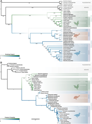
Figure 13. An artistic reconstruction of the skull of Genyornis newtoni, based on all available fossil material, left lateral view. Illustration by Jacob C. Blokland. Scale bar is equal to 50 mm.
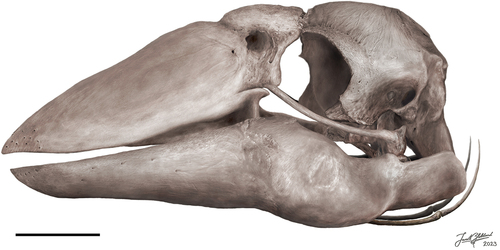
Figure 14. Two select morphological features which show variation across Galloanserae, and, in isolation as single character studies, indicate different phylogenetic hypotheses for the evolutionary placement of the Dromornithidae: A. the presence of either the rostromedial or caudomedial foramen (dark orange) in relation to the crista medialis (light orange) on the quadrate, in medial view; B. the relationship between the aponeurosis zygomatica and processus postorbitalis, in addition to the presence of an ossified portion (dark orange) of the aponeurosis zygomatica (light orange). Dotted and dashed branches in B. illustrate alternative hypotheses discussed in text; the unresolved position illustrated by the dotted line denotes the uncertainty of the order of divergence between the anhimids and dromornithids. Positions of Conflicto antarcticus and the Presbyornithidae are based on Tambussi et al. (Citation2019: fig. 14) and Houde et al. (Citation2023: fig. 9B). Nodes do not correlate with time. Not all families within Galloanserae are displayed. Morphologies drawn from photos and figures in literature: A.: Anseres – Anseranas semipalmata (see Elzanowski and Stidham Citation2010: fig. 5C), Anhimidae – Anhima cornuta (see Elzanowski and Stidham Citation2010: fig. 8B), Presbyornithidae – (see Elzanowski and Stidham Citation2010: fig. 5B), Conflicto antarcticus – (Tambussi et al. Citation2019: fig. 6A), Dromornithidae – Genyornis newtoni (NMV P256893), Megapodiidae – Megapodius freycinet (see Elzanowski and Stidham Citation2010: fig. 5A), Phasianidae – Gallus gallus (FUR 119); B.: Anseres – Sarkidiornis melanotos (see Zusi and Livezey Citation2000: figs. 6D, 7D), Dromornithidae – Genyornis newtoni (SAMA P59516), Anhimidae – Chauna torquata (see Zusi and Livezey Citation2000: figs. 6C, 7B), Megapodiidae – Alectura lathami (SAMA B2439), Phasianidae – Meleagris gallopavo (see Zusi and Livezey Citation2000: fig. 4 G). Silhouettes (designed by PLM) are illustrative representations of species in the same family or appropriate clade, as indicated. Images are not to scale.
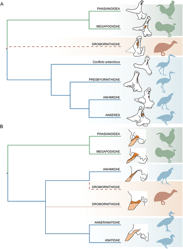
Figure A1. SAMA P 10838, Stirling (Citation1913) original skull of Genyornis newtoni as it is currently: A. lateral view of the skull; B. dorsal view of the skull, showing the block in which the skull has been set. Annotations: ap.zyg.oss., aponeurosis zygomatica ossificans; cond.oc, condylus occipitalis; cr.or., crista orbitalis; pr.post., processus postorbitalis. Image a credit: Mahala Fergusen. Scale bar: 2 cm.
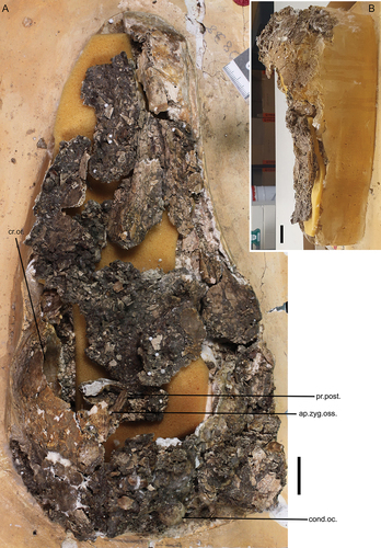
Figure A2. Laterally compressed rostrum, specimen SAMA P59517, transected by a fault in the clay matrix which has offset the caudal part from that more rostral: A. right lateral; B. left lateral; C. dorsal; D. ventral. Annotations: ang.cl., angulus caudolateralis; ca., casque; cr.tom., crista tomialis; cul., culmen; margo.lat., margo lateralis; nas., apertura nasi ossea; pala., os palatinum. Scale bars: 20 mm.
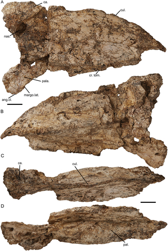
Figure A3. Additional skull specimens of Genyornis newtoni: A. NMV P256893, partial dorsal calvaria in dorsal and ventral views; B. SAMA P53830 jugal arch, right side, medial, lateral, and dorsal views; C. SAMA P53830, right quadrate; D. SAMA P53830 partial condylus occipitalis and occipital region of the cranium; E. SAMA P59520, ramus mandibulae, pars symphysialis, right side, dorsal and ventral views; F. SAMA P59520, partial left lateral cranium in medial (left) and lateral (right) views. Annotations: an.tymp., annulus tympanicus; c. quad, condylus quadratica; cap.s-o., capitulum squamoso-otica; cond.oc., condylus occipitalis; cr.med., crista medialis; cra., cranium; d.rost., depressio rostromedialis; m.a.e., osseous meatus acusticus externus; mand., mandible; quad., quadrate. Scale bars: 10 mm.
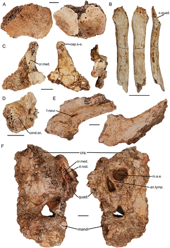
Figure A4. Additional specimens of Genyornis newtoni: A. NMV P256893 partial left skull medial view; B. – G. SAMA P59517, fragmentary mandibular rami, B. right ramus mandibulae, pars caudalis and pars intermedia in lateral view; C. and medial view; D. left ramus mandibulae, pars caudalis et intermedius, lateral view; E. and medial view; F. fragmentary right ramus mandibulae, pars caudalis, dorsal view; G. and rostral view. Abbreviations: c.med., cotyla medialis mandibulae; cer., ceratobranchial; d.rost., depressio rostromedialis; imp.cor., impressio m. AME profundus, pars coronoideus; m.pt.dor., insertion area of m. pterygoideus dorsalis; p.lat., processus lateralis mandibulae; p.med., processus medialis mandibulae; t.prae., tuberculum praearticulare. Scale bar: 10 mm.

Table A1. Genyornis newtoni skull measurements. Rounded to the first decimal point.
Table A2. (Part 1) Homological correlations for the terminology associated with musculus adductor mandibulae across examples of relevant literature.
Supplemental Material
Download Zip (866.6 KB)Data availability statement
All data generated or analysed during this study are included in this published article, the appendices and supplementary information files.