Figures & data
Figure 1. Cretaceous deposits from Brazil and their dinosaur assemblages, emphasising occurrences from the Lower Cretaceous units. 1–3, Botucatu Formation (Paraná Basin); 4–5, Goio Erê and Rio Paraná formations (Caiuá Group); 6–8, Abaeté, Quiricó, and Três Barras formations (Sanfranciscana Basin); 9–10, Maceió and Feliz Deserto formations (Sergipe-Alagoas Basin); 11, Antenor Navarro, Sousa, and Rio Piranhas formations (Rio do Peixe basins); 12, Quixoá (Icó) and Malhada Vermelha formations (Iguatu basins); 13, Rio da Batateira, Crato, and Romualdo formations (Araripe Basin); 14, Açu Formation (Potiguar Basin); 15, Itapecuru Formation (Parnaíba Basin); 16, Alcântara Formation (São Luís-Grajaú Basin). The asterisk exhibits the occurrences from the Recôncavo Basin presented by this work. Detailed information on each occurrence is provided in the Supporting Information (File S1). Modified from ‘Carta Geológica do Brasil ao Milionésimo’ (Souza et al. Citation2004).
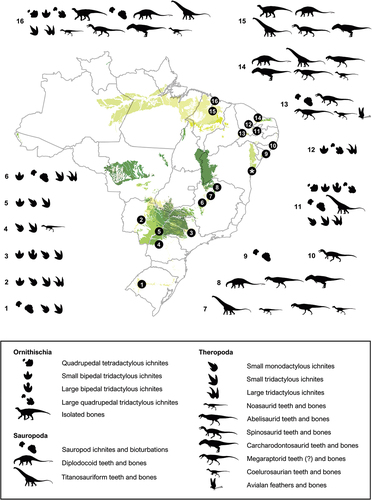
Figure 2. Historical fossil-bearing localities of the Lower Cretaceous Recôncavo Basin in Northeast Brazil, modified from Allport (Citation1860) and Mawson and Woodward (Citation1907). Localities which have yielded dinosaur remains are described in Table 1. Geographic range of the geological units was taken from ‘Carta Geológica do Brasil ao Milionésimo’ (Souza et al. Citation2004). Scale bar = 25 km.
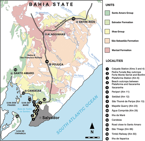
Figure 3. Stratigraphic chart of the Lower Cretaceous Massacará and Ilhas groups (Recôncavo Basin), highlighting their vertebrate assemblages (coloured boxes). 1, hybodontiforms (Acrodus nitidus); 2, semionotids (‘Lepidotes’ spp.); 3, mawsoniids (Mawsonia gigas); 4, undetermined testudines; 5, pholidosaurids (Sarcosuchus hartti); 6, putative gavialoids (‘Thoracosaurus bahiaensis’); 7, pterosaurs (anhanguerids); 8, elasmarians (Tietasaura derbyiana gen. et sp. nov.); 9, iguanodontians; 10, diplodocoids; 11, spinosaurids; 12, carcharodontosaurians; 13, undetermined small theropods (tyrannosauroids?); 14, titanosaurs (lithostrotians); 15, amiids (Calamopleurus mawsoni); 16, cladocyclids (Cladocyclus mawsoni); 17, aspidorhynchids (‘Belonostomus’ carinatus); 18, undetermined clupeomorphs (Scrombroclupeoides scutata); 19, ellimmichthyiforms (Ellimmichthys longicostatum, Ellimmichthys spinosus, Scutatuspinosus itapagipensis). References regarding the vertebrate occurrences were taken through bibliographic survey (see text). Geological chart modified from Da Silva et al. (Citation2007). Silhouettes from several sources.
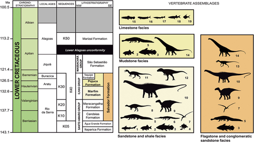
Table 1. Collection details of the specimens described in this work.
Figure 4. Holotypic distal half of the left femur of Tietasaura derbyiana gen. et. sp. nov. (NHM-PV R.3424) from the Valanginian–Hauterivian Marfim Formation (Ilhas Group) at Plataforma Station (Locality 3). A, anterior; B, medial; C, posterior; D, lateral; E, distal views. Anatomical abbreviations: ali, anterior linea muscularis; cd, condylid process; cr, crest; fc, fibular condyle; gr, groove; ieg, intracondylar extensor groove; iff, intracondylar flexor fossa; lfs, lateral fossa; rg, rugosity; sc, sulcus; sf, spiral fracture; scr, supracondylar ridge; st, striae; tc, tibial condyle. Scale bar = 100 mm.
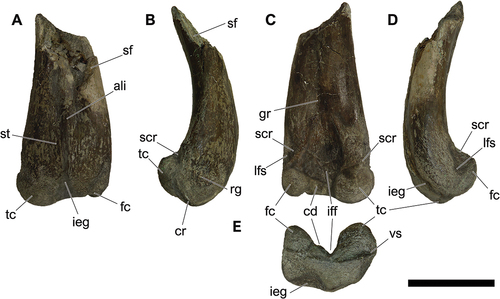
Figure 5. Schematic draw comparing selected elasmarian femora in (1) posterior and (2) distal views. A, Tietasaura derbyiana gen. et. sp. nov. (NHM-PV R.3424); B, Notohypsilophodon comodorensis (UNPSJB-Pv 942); C, Gasparinisaura cincosaltensis (MUCPv-208); D, Fulgurotherium australe (NHM-PV R.3719); E, Trinisaura santamartensis (MLP 08-III-1-1); F, Morrosaurus antarcticus (MACN-Pv 197); G, Isasicursor santacrucensis (MPM 21,533); H, Anabisetia saldiviai (MCF-PVPH 74). Red arrows exhibit the taxa that exhibits a distinct condylid processes between the condyles, while the blue arrows indicate the development and shifting of the condyloid (‘rectangular’) processes. Dotted lines highlight different patterns on general shape (convex or almost rectilinear) and condyle development of femoral surfaces in the posterior (1) and distal (2) views. Anatomical abbreviations: id, indentation; ieg, intracondylar extensor groove. Without scale for comparative purposes.
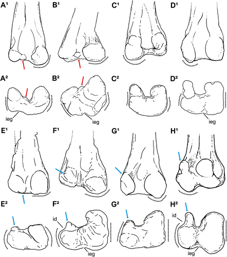
Figure 6. NHM-PV R.3425, isolated middle dorsal centrum with iguanodontian affinities (Specimen A) from the Valanginian–Hauterivian Marfim Formation (Ilhas Group) at Plataforma Station (Locality 3). A, right lateral; B, left lateral; C, dorsal; D, ventral; E, anterior; F, posterior views. Anatomical abbreviations: avh, anteroventral heel; ct, cotyle; nvf, neurovascular foramina; mco, median concavity; ncj, neurocentral joint; pas, posterior articulation surface; pvg, posteroventral groove. Scale bar = 100 mm.
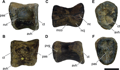
Figure 7. NHM-PV R.2459, isolated middle to posterior dorsal centrum with iguanodontian affinities (Specimen B) from the Valanginian–Hauterivian Marfim Formation (Ilhas Group) at Forte Monte Serrat (Locality 2). A, right lateral; B, left lateral; C, dorsal; D, ventral; E, anterior; F, posterior views. Anatomical abbreviations: avh, anteroventral heel; ct, cotyle; nvf, neurovascular foramina; na, neural arch; nc, neural canal; ncj, neurocentral joint; pas, posterior articulation surface; st, striae. Scale bar = 100 mm.
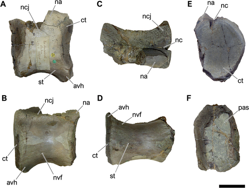
Figure 8. NHM-PV R.3426, middle caudal centrum with iguanodontian affinities (Specimen C) from the Berriasian–Barremian Salvador Formation (Massacará Group) at Mapelle Quarry (Locality 9). A, right lateral; B, left lateral; C, anterior; D, posterior; E, ventral views. Anatomical abbreviations: avh, anteroventral heel; ct, cotyle; pas, posterior articulation surface; pvh, posteroventral heel. Scale bar = 100 mm.
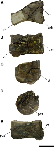
Figure 9. NHM-PV R.3428(a) and NHM-PV R.3428(b), associated middle to posterior caudal centra with iguanodontian affinities (specimen C) from the Berriasian–Barremian Salvador Formation (Massacará Group) at Mapelle Quarry (Locality 9). A-G, right lateral; B-H, left lateral; C-I, dorsal; D-J, ventral; E-K, anterior; F-L, posterior views. Anatomical abbreviations: aas, anterior articulation surface; ct, cotyle; na, neural arch; nc, neural canal; ncj, neurocentral joint; pas, posterior articulation surface; vg, ventral groove. Scale bar = 100 mm.
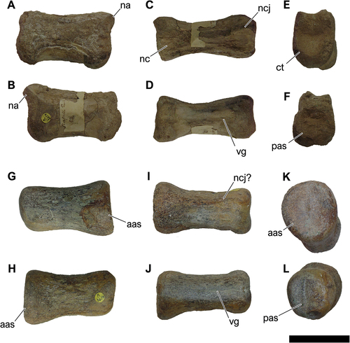
Figure 10. NHM-PV R.3429, isolated middle caudal vertebra with iguanodontian affinities (Specimen D) from the Valanginian–Hauterivian Marfim Formation (Ilhas Group) at Plataforma Station (Locality 3). A, right lateral; B, left lateral; C, dorsal; D, ventral; E, anterior; F, posterior views. Anatomical abbreviations: alf, anterolateral fossa; avh, anteroventral heel; ct, cotyle; ns, neural spine; pas, posterior articulation surface; prz, prezygapophysis. Scale bar = 100 mm.
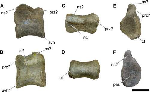
Figure 11. NHM-PV R.2981(b), fragment of a distal middle or proximal posterior caudal centrum with diplodocid affinities (Specimen A) from the Valanginian–Hauterivian Marfim Formation (Ilhas Group) at beach between Plataforma and Itacaranha (Locality 4). A, right lateral; B, left lateral; C, dorsal; D, ventral; E, anterior; F, posterior views. Anatomical abbreviations: bf, blind fossa; na, neural arch; pdc, posterodorsal concavity; pas, posterior articulation surface; pdc, posterodorsal concavity; vh, ventral hollow. Scale bar = 100 mm.
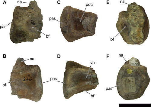
Table 2. Caudal centrum ratios (width-to-height, length-to-height) for selected sauropod taxa (Modified from Tschopp et al. Citation2015: table S39).
Figure 12. NHM-PV R.3427, isolated posterior caudal centrum with diplodocid affinities (Specimen B) from the Berriasian–Barremian Salvador Formation (Massacará Group) at Mapelle Quarry (Locality 9). A, right lateral; B, left lateral; C, dorsal; D, ventral; E, anterior; F, posterior views. Anatomical abbreviations: aas, anterior articular surface; bf, blind fossa; na, neural arch; ncj, neurocentral joint; pdc, posterodorsal concavity; pas, posterior articulation surface; pdc, posterodorsal concavity; rm, rock matrix; vlr, ventrolateral ridge; vh, ventral hollow. Scale bar = 50 mm.
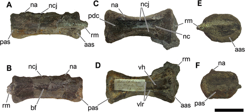
Figure 13. NHM-PV R.2129(a), isolated middle caudal centrum with lithostrotian affinities from the Hauterivian–Barremian Pojuca Formation at Itacaranha (Locality 5). A, right lateral; B, left lateral; C, dorsal; D, ventral; E, anterior; F, posterior views. Anatomical abbreviations: cd, condyle; ce, condylar edge; cf, chevron facet; ct, cotyle; cvj, costovertebral joint; lco, lateral concavity; nc, neural canal; ncj, neurocentral joint; vh, ventral hollow; vlr, ventrolateral ridge. Scale bar = 100 mm.
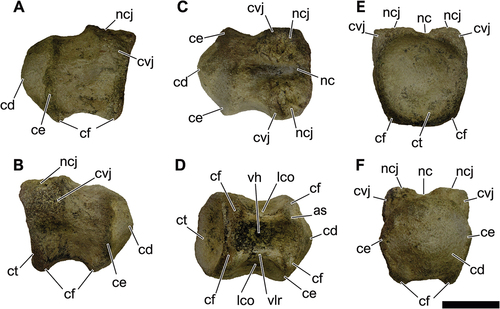
Figure 14. NHM-PV R.2982, right pedal phalanx IV-1 of an indeterminate averostran from the Valanginian–Hauterivian Marfim Formation (Ilhas Group) at beach between Plataforma and Itacaranha (Locality 4). A, dorsal; B, right lateral; C, ventral; D, left lateral; E, anterior; F, posterior views. Anatomical abbreviations: clp, colateral pits; elp, extensor ligament pit; flt, flexor ligament tubercle; gg, ginglymus; pvh, posteroventral heel; tb, tubercle. Scale bar = 50 mm.
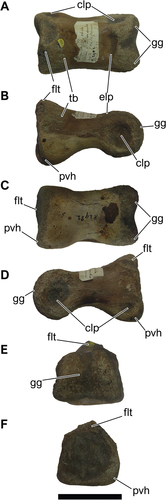
Figure 15. NHM-PV R.2980, anterior caudal centrum with putative spinosaurid affinities from the Valanginian–Hauterivian Marfim Formation (Ilhas Group) at beach between Plataforma and Itacaranha (Locality 4). A, right lateral; B, left lateral; C, dorsal; D, ventral; E, anterior; F, posterior views. Anatomical abbreviations: as, anterior articular surface; bf, blind fossa; cvj, costovertebral joint; nc, neural canal; ncj, neurocentral joint; pas, posterior articulation surface; pvh, posteroventral heel; vh, ventral hollow; vlr, ventrolateral ridge. Scale bar = 100 mm.
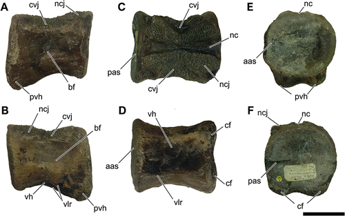
Figure 16. NHM-PV R.2981(a), isolated partial dorsal centrum with putative carcharodontosaurian affinities (Specimen A) from the Valanginian–Hauterivian Marfim Formation (Ilhas Group) at beach between Plataforma and Itacaranha (Locality 4). A, right lateral; B, left lateral; C, dorsal; D, ventral; E, anterior; F, posterior views. Anatomical abbreviations: aas, anterior articular surface; aae, articulation expasion; bf, blind fossa; nc, neural canal; ncj, neurocentral joint; ipt, internal pneumatic tissue. Scale bar = 100 mm.
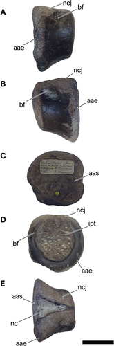
Figure 17. NHM-PV R.2129(b), isolated partial middle caudal centrum with putative carcharodontosaurian affinities (Specimen B) from the Valanginian–Hauterivian Marfim Formation (Ilhas Group) at Itacaranha (Locality 5). A, right lateral; B, left lateral; C, dorsal; D, ventral; E, anterior; F, posterior views. Anatomical abbreviations: aas, anterior articular surface; aae, articulation expasion; mc, medial constriction; pae, posterior articular edge. Scale bar = 100 mm.
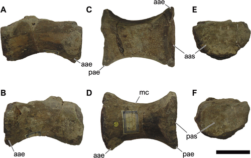
Figure 18. Additional dinosaur specimens from Bahia state (modified from Gallo Citation1993; Gallo et al. Citation2023). A, partial neosauropod tooth crowns (1–3); B, probable theropod humerus with tyrannosauroid affinities in anterior view. Anatomical abbreviations: ap, apical portion; ec, epicondyle; le, laterodistal expansion; ls, labial surface; sm, sinuous margin. Scale bar = 10 mm.
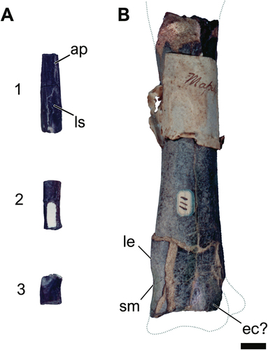
Figure 19. Time-scaled strict consensus trees depicting the phylogenetic relationships of the Recôncavo Basin ornithischians. A, all taxa included in an updated version of the neornithischian phylogenetic matrix of Rozadilla et al. (Citation2019), with Tietasaura derbyiana Gen. et sp. nov. found as an elasmarian, while the material of the Marfim and Salvador formations were recuperated as basal styracosternans. B, phylogenetic affinities of the Marfim and Salvador formations specimens in the phylogenetic matrix of Verdú et al. (Citation2015), which is focused on internal relationships of Iguanodontia. Bremer supports of both analyses are summarised in the supplementary data. Silhouettes from several sources.
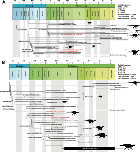
Table 3. Main occurrences of ornithischian bones from South America.
Figure 20. Time-scaled strict consensus tree depicting the phylogenetic relationships of NHM-PV R.2129(a) within Titanosauria, included in an updated version of the matrix of Navarro et al. (Citation2022). Bremer supports are summarised in the supplementary data. Silhouettes from several sources.
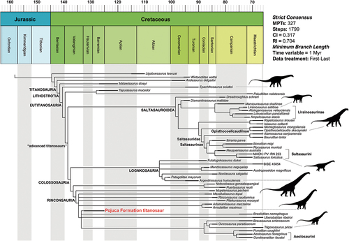
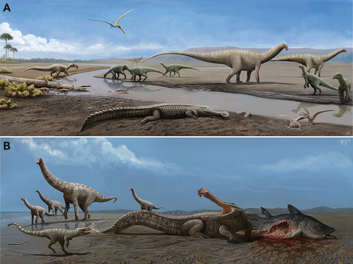
Figure 21. Paleontographical reconstruction of the hypothetical paleoenvironment of the Recôncavo Basin during Lower Cretaceous: A, faunal components of the Marfim Formation (Valanginian–Hauterivian); B, faunal components of the Pojuca Formation (Hauterivian–Barremian). The Salvador Formation is partially synchronous with both units, sharing coeval components. Artwork by Matheus Gadelha.