Figures & data
Figure 1. Location map of the site of the discovery of the holotype of Titanomachya gimenezi. Modified from Sterli et al. (Citation2022)
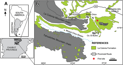
Figure 2. Geological columns of the La Colonia Formation, at Norte de Cerro Bayo, the area of the discovery of the holotype of Titanomachya gimenezi. Modified from Sterli et al. (Citation2022)
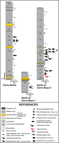
Figure 3. Skeletal reconstruction of Titanomachya gimenezi. Preserved bones shown in light blue. Reconstruction by Gabriel Lio.
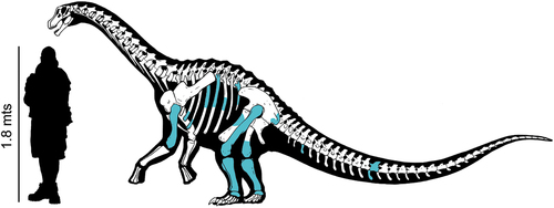
Figure 4. Spatial distribution of Titanomachya gimenezi skeletal elements.
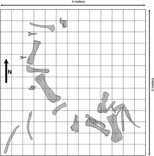
Figure 5. Titanomachya gimenezi, holotype. MPEF Pv 11547/10. Posterior caudal vertebra in A, posterior; B, right lateral; C, anterior; D, dorsal and E, ventral views. Abbreviations: lr, longitudinal ridge. Scale bar = 5 cm.
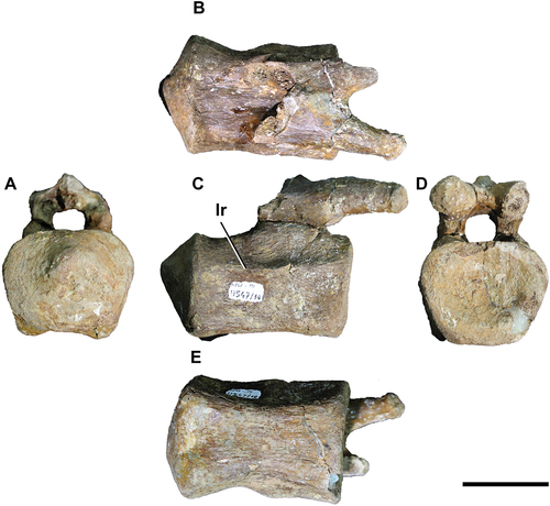
Table 1. Measurements of the elements of the axial skeleton of Titanomachya gimenezi.
Figure 6. Titanomachya gimenezi, holotype. MPEF pv 11547/11 and 12. Middle haemal arch A, right lateral; B, anterior views. Anterior haemal arch C, right lateral; D, anterior views. Scale bar = 10 cm.
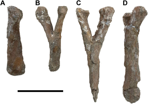
Figure 7. Titanomachya gimenezi, holotype. MPEF Pv 11547/14/1–6. Thoracic ribs fragments. MPEF Pv 11547/1 in A, external and B, internal views. MPEF Pv 11547/2 in C, external view. MPEF Pv 11547/3 in D, external and E, internal views. MPEF Pv 11547/4 in F, external and G, internal views, MPEF Pv 11547/5 in H, external and I, internal views, MPEF Pv 11547/6 in J, internal; K, proximal; L, anterior; M, external and N, posterior views. Scale bar = 30 cm.
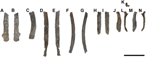
Figure 8. Titanomachya gimenezi, holotype. MPEF Pv 11547/7, left humerus in A, posterior; B, proximal; C,distal; D, medial and E, anterior views. Abbreviations: cb, coracobrachialis origin scar; dc, deltopectoral crest; hh, humeral head; lc, lateral condyle; ld, latissimus dorsi insertion scar; mc, medial condyle; scf, supracondylar fossa; suc, supracoracoid insertion scar. Scale bar = 10 cm.
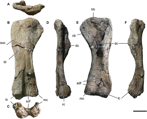
Table 2. Measurements of the forelimb of Titanomachya gimenezi.
Table 3. Measurements of femora and tibiae of Titanomachya gimenezi.
Figure 9. Titanomachya gimenezi, holotype. MPEF Pv 11547/15, left ilia fragment in A, external and B, internal views. Abbreviations: isped, ischial peduncle; poap, postacetabular process. Scale bar = 15 cm.
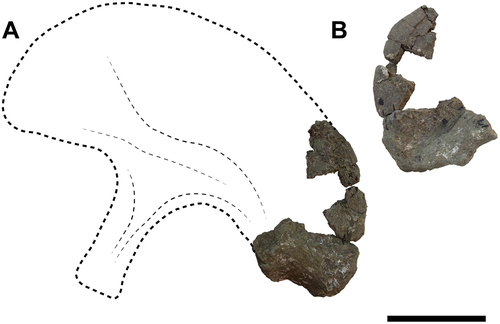
Figure 10. Titanomachya gimenezi, holotype. MPEF Pv 11547/13, indeterminated pube fragment in external view. Scale bar = 20 cm.
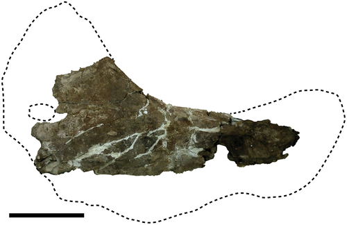
Figure 11. Titanomachya gimenezi, holotype. MPEF Pv 11547/1, right femur in A, anterior; B, proximal; C, medial; D, posterior and E, distal views. Abbreviations: fh, femoral head; gt, greater trochanter; lb, lateral bulge; lec, lateral epicondyle; lc, lateral condyle; mc, medial condyle; pr, posterior ridge; 4t, fourth trochanter. Scale bar = 20 cm.
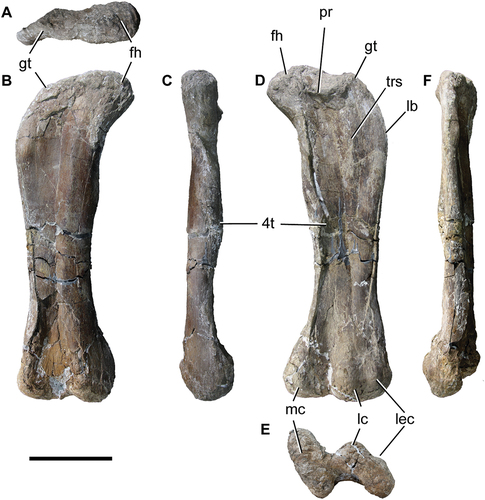
Figure 12. Titanomachya gimenezi, holotype. Tibiae: (a-d) left tibia MPEF 11547/6 in A, anterior; B, distal; C, lateral and D, proximal views. Abbreviations: cc, cnemial crest; cf, cnemial fossa; lc, lateral condyle; pc, posterior condyle. Scale bar = 10 cm.
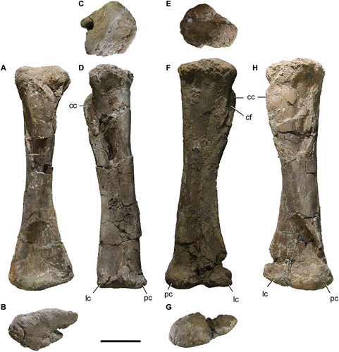
Figure 13. Titanomachya gimenezi, holotype. Fibulae. MPEF 11547/5 left fibula in A, lateral; B, distal; C, proximal, and D, posterior views. MPEF 11547/3 right fibula in E, medial; F, proximal and G, lateral views. Abbreviations: lt, lateral tuberosity. Scale bar = 10 cm.
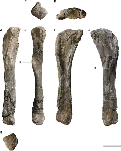
Table 4. Measurements of fibulae and astragalus of Titanomachya gimenezi.
Figure 14. Titanomachya gimenezi, holotype. MPEF 11547/9. Left astragalus in A, anterior; B, dorsal; C, posterior; D, ventral; E, fibular and F, medial views. asp, ascending process. Scale bar = 5 cm.
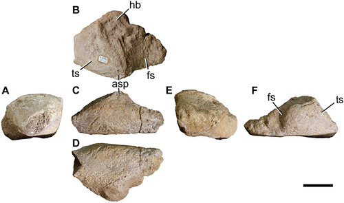
Figure 15. Titanomachya gimenezi, holotype. MPEF 11547/8. Right astragalus in A, anterior; B, dorsal; C, posterior; D, ventral; E, fibular and F, tibial views. Abbreviations: asp, ascending process; fs, fibular surface; hb, hemispherical bulge; ts, tibial surface. Scale bar = 5 cm.
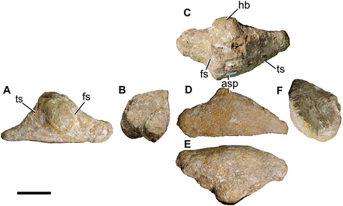
Figure 16. Strict consensus of the phylogenetic analysis. Matrix based on that of Pérez Moreno et al. (Citation2023).
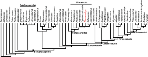
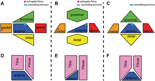
Figure 17. Schematic drawings of the morphologies of astragalus within Titanosauria in all its views. A, diagram of the typical colossosaurianian astragalus and its articulation with tibia and fibula. B, diagram of the typical saltasaurioid astragalus and its articulation with tibia and fibula. C, diagram of the astragalus of Titanomachya and its articulation with the tibia and fibula.