Figures & data
Figure 1. Enhanced Mott cell formation in FcµR-deficient mice. Middle: Mott cells in splenic and lymph node (not shown) tissues from three different strains of Fcmr KO mice as defined by their strong purple staining with PAS reagent are significantly increased as compared with their WT control mice. Bottom: Splenic CD5+B220+ B-1a B cells (106 cells) enriched from Fcmr KO and WT control mice are activated with LPS before fusing with Ag8.653 cells (Ig non-producing plasmacytoma). One IgMλ+ Mott hybridoma clone containing Russel bodies (green) is generated from the total 16 Fcmr-deficient hybridomas, whereas none from the total 6 WT hybridomas. When FcµR cDNA is transduced into the Mott hybridoma, the number of cells containing IgM-inclusion bodies is reduced, the secretion of pentameric IgM (green) in culture media is increased, and secreted IgM binds FcµR (blue) expressed on the plasma membrane. None of these changes are observed when the empty vector as a control is transduced into the Mott hybridoma (not shown). Top: Splenic B cells from Fcmr KO and WT mice are stimulated for 4 d with LPS alone, B-1 (LPS/dextran-anti-IgD/IL-4/IL-5) or B-2 cocktail (anti-CD40/dextran-anti-IgD/IL-4/IL-5). Mott cells are clearly generated in the mutant B cell cultures than the WT control B cell cultures when stimulated with LPS alone or the B-1 stimulation cocktail, but not with the B-2 stimulation cocktail.
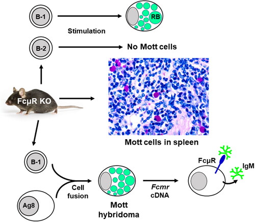
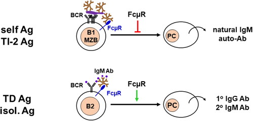
Figure 2. Dysregulated antibody responses. FcµR plays a distinct regulatory role in immune responses depending on the forms of antigens. For cell corpse containing self-antigens, particulate bacteria-associated polysaccharides, and proteins, or TI type2 antigens, FcµR inhibits their antibody reposes as the consequence of multiple BCR cross-linkages (top). On the other hand, for isolated proteins or TD antigens, FcµR enhances the antibody responses as the result of a limited number of BCR cross-linkages.