Figures & data
Figure 1. Topographical SEM images of: (a) and (b) Cicadetta oldfieldi (top and cross-section, respectively) and, (c) and (d) black coloured region of Gudanga sp. nr adamsi (Black cicada) wing membrane (top and cross-section, respectively). (e) Structuring found on the dragonfly (Rhyothemis phyllis chloe) wing membrane. (f) Micron, and (g) sub-micron structures found on a moth wing (Prasinocyma albicosta). (h) and (i) Lacewing (Glenoleon pulchellus) and flower wasp (Scolia soror) nanostructures, respectively, and (j) broad bump structuring of the bladder cicada (Cystosoma schemltzi).
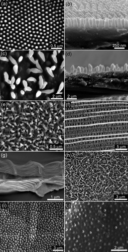
Table 1. Geometrical parameters of cuticle from insect species investigated in this study.
Figure 2. (a) Graph displaying the species type as a function of the apparent contact angle (right axis) and adhesion (left axis). The adhesion measurements were obtained using three different particles (4.5 and 30 μm silica beads, and an AFM silica tip (ca 28 nm)). Error bars represent SEs. Photographs showing a 10 μl droplet deposited on (b) a cicada wing membrane, and (c) hydrophobic PDMS surface. (d) and (e) show the PDMS replica of the cicada Tosena sybilla and the interaction of a water droplet with a polymer (PDMS) replica of the cicada Gaeana cheni, respectively.
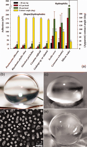
Figure 3. SEM images showing the orientation of the contaminating particulates attached to AFM levers. (a) Pimelea linifolia ssp., (b) Grevillea ‘Red Sunset’, (c) Acacia fimbriata, and (d) 30 μm silica bead.
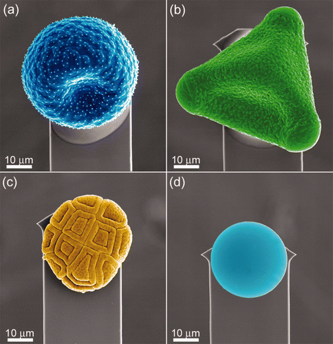
Table 2. Dimensional parameters of all the particles used for adhesion measurements.
Figure 4. Graph displaying the relationship between the apparent contact angle and adhesion with the C18 particles on the hydrophilic and super/hydrophobic insect wing surfaces. Error bars represent SEs.
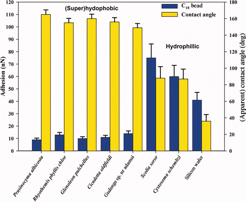
Figure 5. Graph displaying the apparent contact angle and adhesion with the three different pollen particles – ⋄ Pimelea linifolia ssp., ▵ Grevillea ‘Red Sunset’ and ˆ Acacia fimbriata ‘Golden wattle’. Error bars represent SEs.
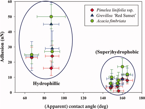
Figure S1. An optical microscope image of a water droplet resting on the wing membrane of a cicada revealing an apparent contact angle (CA) of ca 150° with the surface.
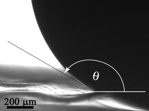
Figure S2. Top (a) and side (b) view, SEM images of a PDMS replica of the black cicada (Gudanga sp.).
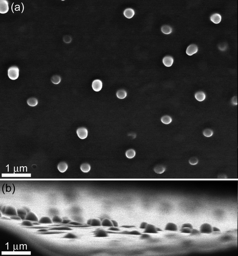
Figure S3. Top and side SEM images of the coloured regions found on the cicada wings of Tosena sybilla ((a) and (b), respectively) and Gaeana cheni ((c) and (d), respectively).
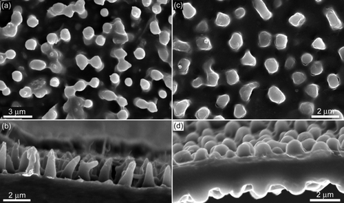
Figure S4. A dehydrated (a) and ‘fresh’ (b) forewing of the bladder cicada, Cystosoma schmeltzi.
Figure S5. SEM images of the finer surface features of the three pollen surfaces used. (a) Pimelea linifolia ssp., (b) Gravillea ‘Red Sunset’ and (c) Acacia fimbriata.
Figure S6. AFM images of a silica bead, revealing the finer surface structures. (a) A topographical image, (b) a 3-d representation, and (c) an artificially shaded and leveled 3-d image showing the fine surface features. The imaging was performed with the bead attached to a lever and scanned over a calibration grid, ie reverse imaging.
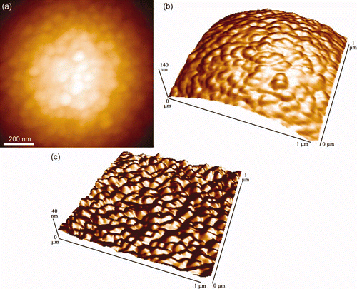
Figure S7. A single representative f-d curve showing multiple ‘jumps’ on the retract curve (red) obtained on the moth wing (Prasinocyma albicosta). The approach curve (blue) shows a distinct snap-on feature (and a general downward trend) where the scales are presumably statically attracted to the surface of the Si bead.
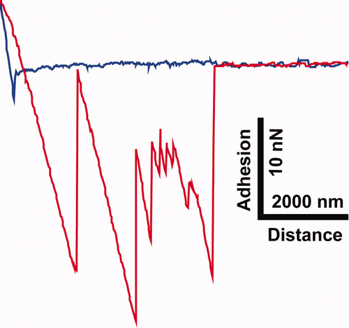
Figure S8. The overlapping scale arrangement of the Prasinocyma albicosta moth wing.
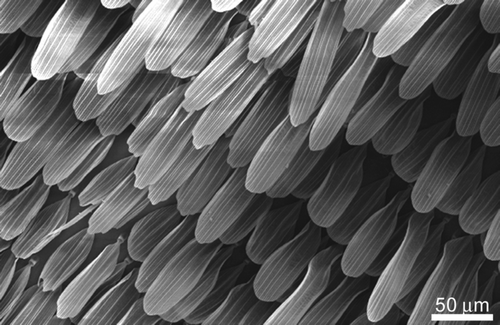
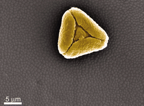
Figure S9. A pollen grain found on the wing membrane of the hydrophilic flower wasp (Scolia soror).