Figures & data
Figure 1. Adherence patterns of H. capsulatum yeast to pneumocytes. Immunofluorescence staining of non-infected pneumocytes (A). Infection after incubation for 7 h with the EH-315 strain in pneumocytes (B) and the 60I strain in pneumocytes (C). Phalloidin-FITC (green); DAPI (blue); anti-Histoplasma polyclonal serum plus Alexa 594-conjugated antibody (red-to-yellow). The assays were conducted in an IN Cell Analyzer 2000 using light microscopy.
Figure 2. Kinetics of biofilm formation by H. capsulatum on polystyrene microtiter plates. The two H. capsulatum strains were processed by the colorimetric XTT reduction assay as described in Materials and methods. The measurement average data of three XTT assays was considered to generate the figure. The assay was performed twice, with similar results. P values of <0.05 were considered significant.
Figure 3. Microphotograph taken using a camera attached to an inverted microscope of the kinetics of biofilm formation by H. capsulatum in polystyrene microtiter plates after aspiration of BHI medium and subsequent washings with PBS, as determined by the colorimetric XTT reduction assay. The average of three XTT assay measurements was taken. This experiment was performed twice, with similar results each time. Bars = 100 μm for panels A, B, C, and D.
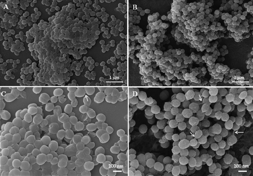
Figure 4. SEM images of mature biofilms of the EH-315 Mexican bat strain of H. capsulatum formed after incubation for 72 h at 37°C (A, C) and of the 60I Brazilian clinical strain of H. capsulatum also formed after incubation for 72 h at 37°C (B, D). Arrows denote yeasts cells bound by an exopolymeric matrix.