Figures & data
Table 1. Materials, compounds, and the components used in the present study.
Figure 1 Flow-chart of the experimental design of the study.
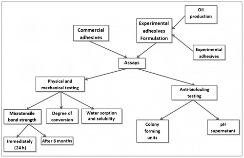
Figure 2 Mean viable bacteria (CFU cm−2 dry biofilm weight) in biofilms grown for 72 h (n = 10). The data were normalized by transforming by log10. Within a panel, group values that were identified using similar lower case letters were not significantly different (p > 0.05). A = total microorganisms; B = total aciduric bacteria; C = S. mutans; D = total lactobacilli bacteria.
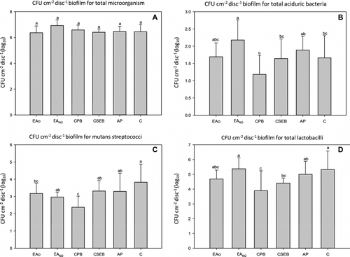
Figure 3 Chromatograms of the fatty acid profile extracted from seeds and standard fatty acids, respectively, of B.capitata.
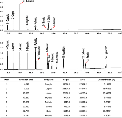
Table 2. Supernatant pH (mean ± SD) of the storage medium of each adhesive system.
Table 3. Microtensile bond strength (MPa), at different times of storage (mean ± SD).
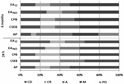
Figure 4 Distribution of failure modes among the groups. CD = cohesive failure within dentin; CR = cohesive failure within resin; A = adhesive failure; M = mixed failure; PD = prematurely debonded specimen. For each group and evaluation period n = 20.