Figures & data
Figure 1 Combined treatment of P. aeruginosa biofilms with lactoferrin and xylitol results in membrane disruption. SEM imaging (panels A and B) and LIVE/DEAD staining (panels C and D) demonstrate significant membrane disruption of treated cells (panels B and D) when compared to untreated cells (panels A and C). For more information see Ammons, Ward and James (Citation2011) and Ammons, Ward, Dowd et al. (Citation2011).
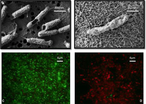
Table 1. Antimicrobial efficacy of lactoferrin and peptide derivatives against MRSA in vitro and in vivo.
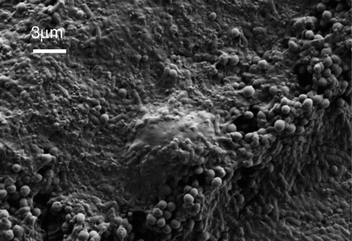
Figure 2 SEM image showing an undifferentiated HL60 human monocytic cell which has come up against a wall of P. aeruginosa and S. aureus cells co-cultured in a biofilm grown in vitro.