Publication Cover
Biofouling
The Journal of Bioadhesion and Biofilm Research
Volume 29, 2013 - Issue 8
2,170
Views
22
CrossRef citations to date
0
Altmetric
Articles

Update on the challenging role of biofilms in peritoneal dialysis

, , , , &
Pages 1015-1027 | Received 07 Mar 2013, Accepted 28 Jun 2013, Published online: 02 Sep 2013

Figures & data

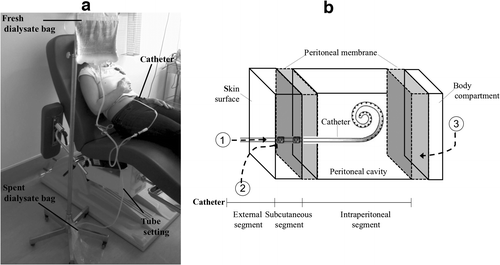
Figure 1 PD technique. (a) A patient is shown during an exchange. The patient is connected to the tubing set and the spent dialysate is drained out. Then, the peritoneal cavity is filled with fresh dialysate and the tubing set is disconnected. After a dwell, the process is repeated. (b) A schematic diagram indicating the relationship between the PD catheter and the adjacent tissues. The potential pathways of peritoneal cavity contamination are indicated: 1-intraluminal, 2-periluminal, and 3-transmural/hematogenous/transvaginal (adapted from Spencer (Citation1988)).

Figure 1 PD technique. (a) A patient is shown during an exchange. The patient is connected to the tubing set and the spent dialysate is drained out. Then, the peritoneal cavity is filled with fresh dialysate and the tubing set is disconnected. After a dwell, the process is repeated. (b) A schematic diagram indicating the relationship between the PD catheter and the adjacent tissues. The potential pathways of peritoneal cavity contamination are indicated: 1-intraluminal, 2-periluminal, and 3-transmural/hematogenous/transvaginal (adapted from Spencer (Citation1988)).

Table 1. Summary of studies carried out on PD catheters supporting the presence of a microbial biofilm.

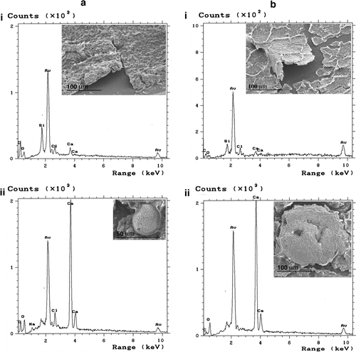
Figure 2 Example of SEM and EDX analyses performed on the intraluminal surface of transfer sets removed from two patients (a and b). Biofilm overview (i) and specific encrustations (ii) observed by SEM are shown as inserts to the corresponding EDX spectrum illustrating the elemental composition. Au (from sputtering-coating of samples) and Si (from the catheter wall) are not elements originating from the biofilms.

Figure 2 Example of SEM and EDX analyses performed on the intraluminal surface of transfer sets removed from two patients (a and b). Biofilm overview (i) and specific encrustations (ii) observed by SEM are shown as inserts to the corresponding EDX spectrum illustrating the elemental composition. Au (from sputtering-coating of samples) and Si (from the catheter wall) are not elements originating from the biofilms.

Table 2. Microbiological findings on PD catheters.
Reprints and Corporate Permissions

Please note: Selecting permissions does not provide access to the full text of the article, please see our help page How do I view content?

To request a reprint or corporate permissions for this article, please click on the relevant link below:

Academic Permissions

Please note: Selecting permissions does not provide access to the full text of the article, please see our help page How do I view content?

Obtain permissions instantly via Rightslink by clicking on the button below:

If you are unable to obtain permissions via Rightslink, please complete and submit this Permissions form. For more information, please visit our Permissions help page.