Figures & data
TABLE 1. Localization of the defects indicating the operation.
FIGURE 1. Photo documentation of the surgical intervention. A: A patient selected for capsuloplasty. B: The dissected capsule flap. C: The implant after positioning and covering with the capsule flap. D: The closed wound. E: The healed wound 3 month after the operation.
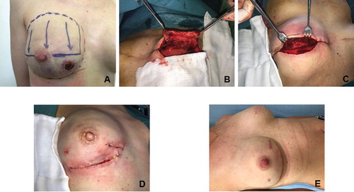
FIGURE 2. The blood flow of capsule flaps and thoraco-epigastrial fasciocutaneous flaps. Caps.: capsulotomy, Inc.: incision, Prep. of the flap: preparation of the flap, Fix. of the flap: fixation of the flap. Values are referred to the baseline and are given as percentage. Median values with 25th and 75th percentiles are demonstrated. A: blood flow of the capsule flaps in different stages of the operation. B: perfusion of the thoraco-epigastrial fasciocutaneous flaps during different steps of the intervention.
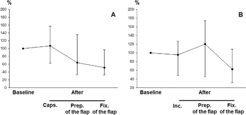
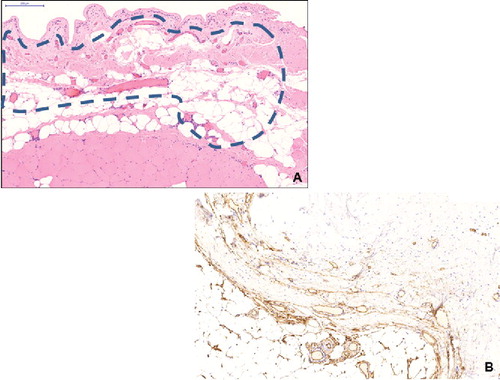
FIGURE 3. A: Low power micrograph of the capsule (haematoxylin-eosin staining, slide scanning, scale bar: 200 μm). Perforating vessels in the connecting tissue between striated muscle and capsule (dashed line). B: CD34-positive structures in the capsule (immunohistochemistry for CD34, counterstaining with haematoxylin, CD34-positive parts appear brown).