
Figures & data
FIGURE 1. Different steps of the operation. A: preoperative status, B: dissection of the subcutaneous and cutaneous pedicled flap, C: temporarily situated flap, blue ink shows the position of alar-facial groove on the skin flap, D: alar-facial groove is fixed with 5/0 absorbable sutures, E: the flap is sutured into the defect, F: late postoperative status.
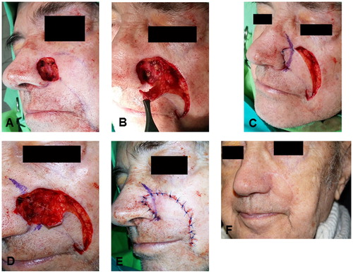
FIGURE 2. Reconstruction in another patient. A: preoperative status, B: preparation of the flap, C: positioning of the flap, D: early postoperative status, E and F: late postoperative status.
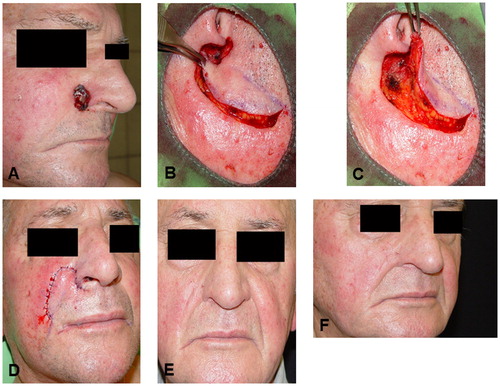
FIGURE 3. Application of the combined flap in a third patient. A: preoperative status, B: dissection of the flap, C: positioning of the flap, white arrow: the subcutaneous part of the flap via which perforators enter the flap, black arrow: direction of the cutaneous branches from the medial part of the flap, D: early postoperative status, E and F: late postoperative status.
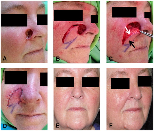
FIGURE 4. A further patient with the combined flap. A: preoperative status, B: preparation of the flap, C: positioning of the flap, D: early postoperative status, E and F: late postoperative status.
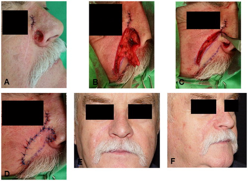
TABLE 1. Frequency and severity of the postoperative complications
FIGURE 5. The blood flow of the flaps before the operation, during the intervention and in the postoperative period. Prep. of the flap: preparation of the flap, Fix. of the flap: fixation of the flap. Median values with 25th and 75th percentiles are demonstrated. X: p < .05 vs. Baseline.
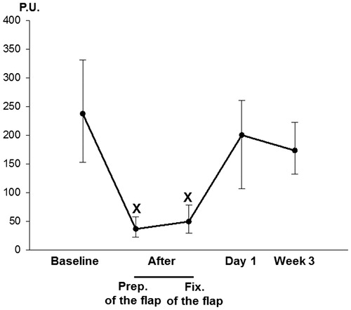
TABLE 2. Patient satisfaction