Figures & data
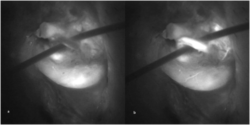
FIGURE 1. Intraoperative indocyanine green angiography was performed after ligation of all internal spermatic veins. (a) Significant quantity of residual indocyanine green signal remained in the field after the previous three angiograms. (b) The strong signal from testicular artery was visualized and clearly differentiated from the background.