
Figures & data
Figure 1. Self-assembled visual needle: (A) needle sheath; (B) Y-adapter #1; (C) fiber optic bundle (fiber endoscope); (D) Y-adapter #2; (E) Irrigation tube; (F) Holmium laser fiber.
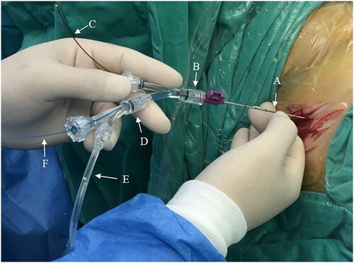
Figure 2. Holmium laser lithotripsy during microperc: (A) puncture needle sheath; (B) Holmium laser fiber; (C) kidney stones.
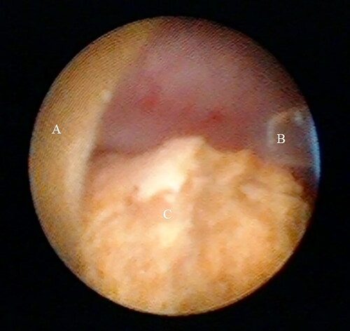
Figure 3. Stone powder and fragments (white arrow) flushed out through the ureteral access sheath (red arrow) during lithotripsy.
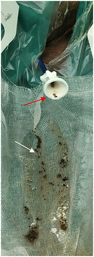
Table 1. Demographic data and stone characteristics.
Figure 4. Pre-and post-operative CT and abdominal plain film (KUB) of left low calyceal stone treated with microperc: (A) preoperative CT; (B) KUB; (C) stone and fragments disappeared on postoperative day 1.
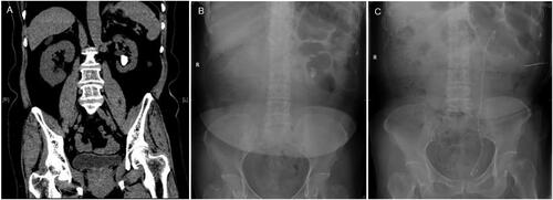
Table 2. Intra-and postoperative Findings.
Availability of data and material
The datasets used and/or analyzed during the current study are available from the corresponding author on reasonable request.