Figures & data
Table 1. Sequences of primers designed for amplification of genes.
Figure 1. Effect of ACT on BMD and bone microstructure in the GIOP rat model. A. ACT significantly reversed BMD in the Dex-induced OP rat model. B–E. Changes in the microarchitectural parameters of bone such as BV/TV, Tb.N, Tb.Th, Tb.Sp in response to ACT treatment and Dex induction. BV/TV, bone volume over total volume; Tb.N, trabecular number; Tb.Th, trabecular thickness; Tb.Sp, trabecular separation; ACT-L, 50 mg/kg/d ACT; ACT-H, 100 mg/kg/d ACT. N = 8, ***P < 0.001 vs. control; ##P < 0.01, ###P < 0.001 vs. Model.
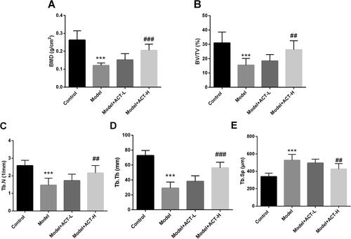
Figure 2. Regulation of bone metabolism in the Dex-induced OP rat model by ACT. A. ALP activity assay kit was used to evaluate serum ALP levels. ELISA was performed to assess the levels of OC (B) and CTX (C) in the serum of rats with GIOP. RT-qPCR was performed to assess the expression of osteogenic differentiation-related genes (D) and bone resorption-related genes (E). N = 8, ***P < 0.001 vs. control; #P < 0.05, ##P < 0.01, ###P < 0.001 vs. Model.
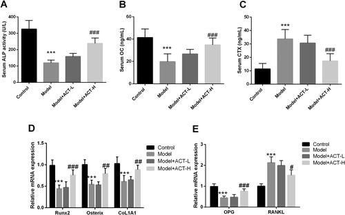
Figure 3. ACT significantly alleviates the effect of Dex on osteoblast activity and apoptosis. Resultst of the CCK-8 assay on Dex (A), ACT (B), and co-treatment with Dex and ACT (C) MC3T3-E1 viability in osteoblasts. D. Flow cytometry assays for the regulation of osteoblast apoptosis by ACT and Dex co-treatment. **P < 0.01, ***P < 0.001 vs. 0 group; ##P < 0.01, ###P < 0.001 vs. Dex 1 μM and 0 M ACT.
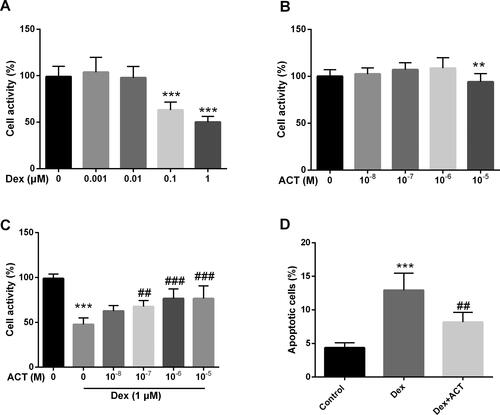
Figure 4. Effects of ACT on Dex-induced bone transforming factor and key proteins of the PI3K/AKT/mTOR signaling pathway in osteoblasts. A. An ALP activity assay kit was employed to evaluate ALP levels in osteoblasts. RT-qPCR analysis of mRNA levels of genes related to osteogenic differentiation (B) and genes related to bone resorption (C). Western-blot analysis (D) and quantification of key protein levels (E) in the PI3K/AKT/mTOR signaling pathway. ***P < 0.001 vs. Control; #P < 0.05, ##P < 0.01, ###P < 0.001 vs. Dex.
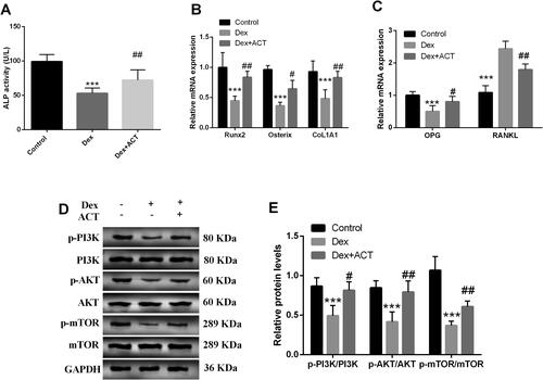
Figure 5. The PI3K inhibitor LY294002 reverses the protective effect of ACT on osteoblast activity andapoptosis. A and B. Western blot analysis of LY294002 action on the ACT-induced PI3K/AKT/mTOR signaling pathway key protein levels and quantification. The CCK-8 assay (C) and flow cytometry (D) to assess the effect of LY294002 on osteoblast activity and apoptosis. ***P < 0.001 vs. Control; #P < 0.05, ##P < 0.01, ###P < 0.001 vs. Dex, & P < 0.05 vs. Dex + ACT.
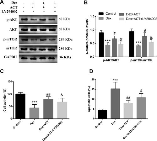
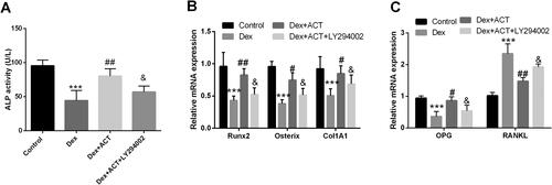
Figure 6. LY294002 reverses the regulation of osteoblast differentiation and bone resorption by ACT. An ALP activity assay kit was used to evaluate ALP levels in osteoblasts. RT-qPCR analysis of the expression of osteoblast differentiation genes and bone resorption-related genes in osteoblasts exposed to Dex and ACT with LY294002. ***P < 0.001 vs. Control; #P < 0.05, ##P < 0.01, ###P < 0.001 vs. Dex; & P < 0.05 vs. Dex + ACT.