Figures & data
Figure 1. CT value measured from pre-operative CT scan. (a,b) lumbar lordosis (LL), (c,d) segmental lumbar lordosis (SLL), (e) Anterior height of the intervertebral space, (f) Posterior height of the intervertebral space, SLL: segment lumbar lordosis, LL: lumbar lordosis.
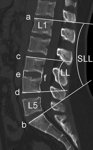
Figure 2. Cage position. The endplate was divided into 4 regions. (a) anterior-inferior edge of the cage falls on zone I. (b) anterior-inferior edge of the cage falls behind zone I.
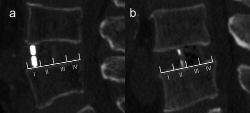
Table 1. Characteristics And intraoperative endplate injury of patients received LLIF or TLIF surgery.
Table 2. Univariate Analysis for endplate injury in LLIF group.
Table 3. Binary logistic regression analysis for endplate injury in LLIF group.
Figure 3. The patterns of intraoperative endplate injury during LLIF procedures. (a) Endplate abrasion, without cage settling. (b) one edge of the cage settles into the vertebral body. (c) two diagonal edges of the cage settle into the vertebral bodies. (d) the upper or lower surface of the cage settles into the vertebral body. (e) Fracture of the superior or inferior vertebral body, the fracture line is distributed along the coronal plane.
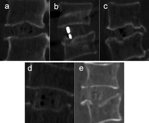
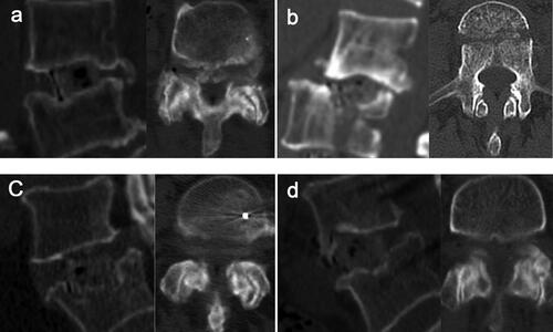
Figure 4. (a)–(b) The anterior-inferior edge of the cage falls on the anterior part of the epiphyseal ring and the cage footprint area on the endplate. (c)–(d): the anterior-inferior edge of the cage falls behind the anterior part of the epiphyseal ring and the cage footprint area on the endplate.
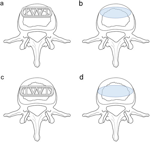
Figure 5. (a)–(d) Severe endplate damage or even coronal vertebral fractures accompanied by facet joint degeneration.