
Figures & data
Figure 1. EGFR mutational status of lung adenocarcinoma (LUAD). Abbreviation: EGFR-mt, EGFR mutation adenocarcinoma; EGFR-wd, EGFR wild type adenocarcinoma.
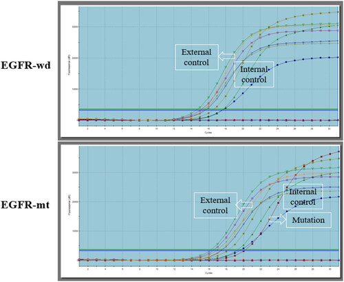
Table 1. The clinicopathological features of 89 LUAD patients.
Figure 2. Different expressions of HIF-1α and PKM2 in normal lung tissue (NLT) and lung adenocarcinoma (LUAD).
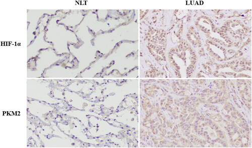
Table 2. Expressions of HIF-1α and PKM2 in normal lung tissue and lung adenocarcinoma.
Table 3. Relationship between the expression of HIF-1α and PKM2 in lung adenocarcinoma.
Table 4. Relationships of HIF-1α/PKM2 feedback loop proteins with clinical pathological characteristics in adenocarcinomas of lung.
Figure 3. Different expressions of HIF-1α and PKM2 in EGFR wild type (EGFR-wd) and EGFR mutation (EGFR-mt) lung adenocarcinomas.
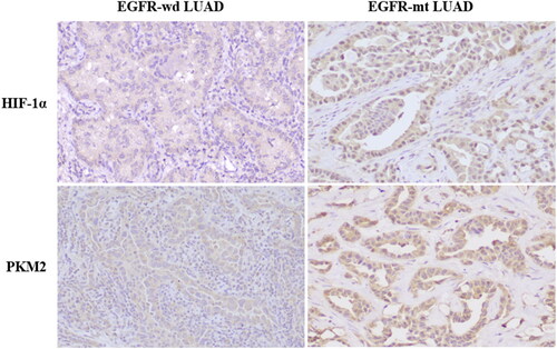
Table 5. HIF-1α and PKM2 protein expressions in EGFR wild-type versus EGFR-mutated lung adenocarcinoma.
Table 6. Relationship between the expression of HIF-1α and PKM2 protein in EGFR wild-type versus EGFR-mutated lung adenocarcinoma.
Table 7. Relationships of HIF-1α/PKM2 feedback loop proteins with clinical pathological characteristics in EGFR wild-type versus EGFR-mutated lung adenocarcinoma.