Publication Cover
Inhalation Toxicology
International Forum for Respiratory Research
Volume 21, 2009 - Issue 1
482
Views
7
CrossRef citations to date
0
Altmetric
Research Article

Toxicogenomic Analysis of Mainstream Tobacco Smoke-Exposed Mice Reveals Repression of Plasminogen Activator Inhibitor-1 Gene in Heart

, , , , &
Pages 78-85 | Received 23 Apr 2008, Accepted 15 May 2008, Published online: 01 Jan 2009

Figures & data

TABLE 1 List of genes up or down regulated in the treatment groups

FIG. 1 Validation of microarray results. Data are presented as fold change over control values (n = 5 mice/group, ± SEM): 1, 6 wk smoke; 2, 6 wk smoke + 6 wk break; 3, 12 wk smoke; and 4, 12 wk smoke + 6 wk break. Asterisk indicates significant at p< .05, t-test conducted on RT-PCR values.
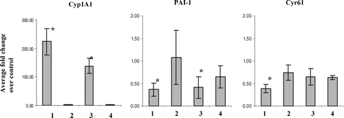
FIG. 1  Validation of microarray results. Data are presented as fold change over control values (n = 5 mice/group, ± SEM): 1, 6 wk smoke; 2, 6 wk smoke + 6 wk break; 3, 12 wk smoke; and 4, 12 wk smoke + 6 wk break. Asterisk indicates significant at p< .05, t-test conducted on RT-PCR values.

FIG. 2 Cyp1A1 protein abundance in heart microsome extract (n= 5). MW: molecular weight marker; +ve control: BaP-treated lung microsome extract. Lanes 1–4: treatment groups, 6 wk smoke, 12 wk smoke, 6 wk smoke + 6 wk break, and 12 wk smoke + 6 wk break; lanes 5–8: matched controls.
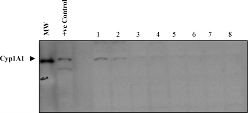
FIG. 2  Cyp1A1 protein abundance in heart microsome extract (n= 5). MW: molecular weight marker; +ve control: BaP-treated lung microsome extract. Lanes 1–4: treatment groups, 6 wk smoke, 12 wk smoke, 6 wk smoke + 6 wk break, and 12 wk smoke + 6 wk break; lanes 5–8: matched controls.

FIG. 3 (A) Total immunoreactive PAI-1 in heart tissue extracts. (B) PAI-1 activity in heart tissue extracts. Data represent fold change over matched controls (n= 5, ± SEM): 1, 6 wk smoke; 2, 6 wk smoke + 6 wk break; 3, 12 wk smoke; 4, 12 wk smoke + 6 wk break.
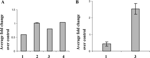
FIG. 3  (A) Total immunoreactive PAI-1 in heart tissue extracts. (B) PAI-1 activity in heart tissue extracts. Data represent fold change over matched controls (n= 5, ± SEM): 1, 6 wk smoke; 2, 6 wk smoke + 6 wk break; 3, 12 wk smoke; 4, 12 wk smoke + 6 wk break.

FIG. 4 (A) Western blot of immunoreactive tPA in heart tissue extracts. Lanes 1, 3, 5, and 7 represent 6 wk smoke, 6 wk smoke + 6 wk break, 12 wk smoke, and 12 wk smoke + 6 wk smoke. Lanes 2, 4, 6, and 8 represent matched controls. (B) Total tPA activity present in heart tissue extracts. Data shown as fold change over matched controls:1, 6 wk smoke; 2, 6 wk smoke + 6 wk break; 3, 12 wk smoke; 4, 12 wk smoke + 6 wk break.

FIG. 4  (A) Western blot of immunoreactive tPA in heart tissue extracts. Lanes 1, 3, 5, and 7 represent 6 wk smoke, 6 wk smoke + 6 wk break, 12 wk smoke, and 12 wk smoke + 6 wk smoke. Lanes 2, 4, 6, and 8 represent matched controls. (B) Total tPA activity present in heart tissue extracts. Data shown as fold change over matched controls:1, 6 wk smoke; 2, 6 wk smoke + 6 wk break; 3, 12 wk smoke; 4, 12 wk smoke + 6 wk break.