
Figures & data
Figure 1. Study design and schedule. Mice were assigned to either depleted/unenriched (DH) or enriched (EH) housing. A subset of animals (cohort A) in each of the housing groups underwent surgical implantation of radiotelemeters during week 3, while the rest (cohort B) remained untelemetered. All the animals were maintained in their respective housing conditions for approximately 6–7 weeks. All cohort animals were exposed to filtered air (FA) during week 5 and eucalyptus wildfire smoke (WS) during week 6, and then monitored for 5–7 days and then necropsied. Cohort B animals underwent ventilatory function assessments after an initial week 5 exposure to FA and then WS during week 6 and were then necropsied 24 h later. A parallel set of animals in cohort B were only exposed to FA in week 6 and necropsied thereafter. (image created in biorender).
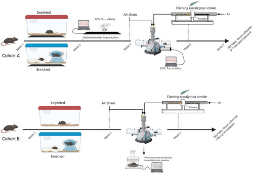
Figure 1. Living conditions alter cardiac gene expression in mice. Volcano plot showing the differences in expression between DH and EH mice. Each dot represents a gene within the comparison performed. The coloring on the dots indicates the clustering information for each gene, those in black are outside the parameters of the filter and not statistically significant. Genes in green were either significantly increased (ACTA, AR, GJA5, HCRTR1, NPPA, NPPB, P75) or decreased (TRPV1) in EH hearts when compared to DH with a 1.25-fold change cutoff. Each are depicted in the box and whisker plot. When the filter was set to 1.15-fold change, additional genes showed significant increases at p < 0.05 (red). n = 11–12; *significantly different from DH, p < 0.5.
Table 1. Exposure details.
Table 2. Heart rate variability before and during eucalyptus smoke exposure.
Figure 2. Living in a depleted housing environment impacts change in body weight. A. There was no difference in body weights at Week 1 when animals were separated into DH (open circles) and EH (filled circles). All the animals, Cohorts A mice experienced an increase in body weight during the course of the study. B. In cohort A, EH mice had less weight gain when compared to DH after radiotelemetry surgery. n = 18–20; * significantly different from DH, p < 0.05.
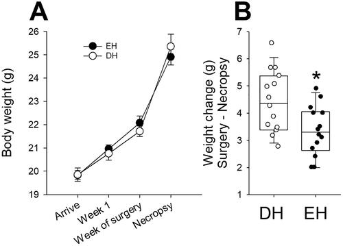
Figure 3. Housing conditions affect core body temperature. A. Core body temperature was normal in Cohort A mice housed in EH (filled circles) after radiotelemetry surgery when compared to DH (open circles), which had a significant decrease. B. There were no core body temperature differences between DH and EH after air sham or WS exposure, however, there was an increasing trend in the former. n = 10–12; * significantly different from DH, p < 0.05.
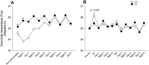
Figure 4. Housing conditions affect day and nighttime heart rate in mice. Although HR was measured continuously after surgery until the end of the study, only data from relevant segments are shown here because the intervening periods had circadian patterns for all animals. A. Heart rate was significantly increased during the nighttime in mice housed in an enriched environment (filled circles) immediately after radiotelemetry surgery when compared to depleted housing (open circles). The heart rates of both groups became comparable thereafter. In addition, mice living in an enriched environment had significantly lower heart rates, especially after the air sham and WS exposures, when compared to depleted housing. B. During the one-hour exposure to WS, mice living in an enriched housing environment had less change in heart rate relative to the air sham exposure and C. significantly fewer non-conducted p-wave or block arrhythmia when compared to depleted mice. D. Representative trace showing electrocardiogram with anon-conducted p-wave. n = 10–12; * significantly different from DH, p < 0.05.
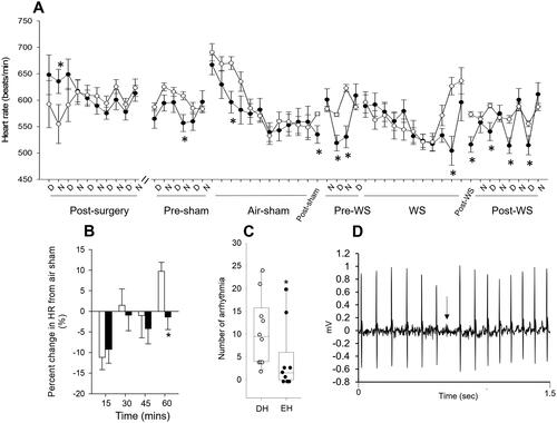
Figure 5. Housing conditions alter activity after air sham and wildfire smoke exposure. All mice experienced blunted activity during the 3 to 4 days after radiotelemeter surgery, but all of them established normal activity thereafter (higher counts/min at night and lower during the day), which continued until the air sham exposure (data shown is not continuous for the entire duration because there was no change). Animals housed in enriched housing (filled circles) had increased activity one day after air sham exposure and immediately after WS when compared to DH (open circles). n = 10–12; * significantly different from DH, p < 0.05.
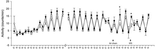
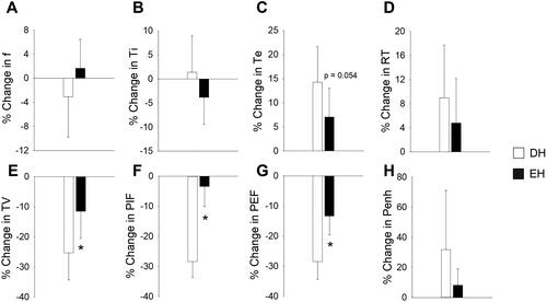
Figure 6. Housing conditions alter the ventilatory response to wildfire smoke exposure. Responses were measured immediately after WS exposure and the percent change from air sham responses were determined. EH mice had significantly less decrease in TV (E), PIF (F), and PEF (G) after WS when compared to DH. There were no other significant differences. n = 8–9; * significantly different from DH, p < 0.05.
Data availability statement
All data are available on EPA ScienceHub.