Figures & data
Figure 1. Three structures of amphotericin B: a chemical drawing showing the hydrophobic (lipophilic) polyene (yellow), and the hydrophilic poly-hydroxyl side (blue); a space filling model; and a model of the putative structure of a 16 membered double barrel channel entrapped in the phospholipid bilayer associated with cholesterol.
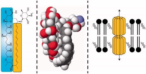
Figure 2. Unlabeled, lyophilized small volume parenteral vials of liposomal amphotericin B (AmBisome).
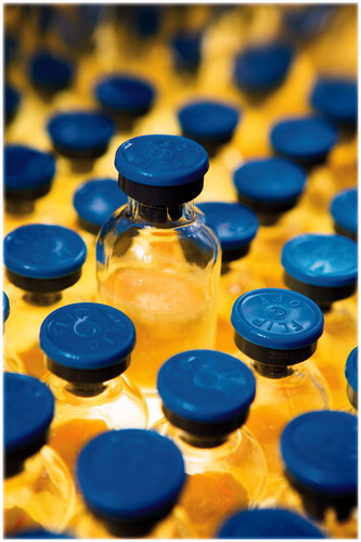
Figure 3. AmBisome diluted in 5% dextrose (D5W) compared to placebo-to-match AmBisome (FMN/SoyPC liposomes).
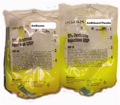
Figure 4. Measured phosphate using Beckman-Coulter PHOSm or PHS kits, spiked with AmBisome or AmBisome placebo (AmBisome formula absent drug). Measurements are untreated, microcentrifuged, or treated with lipospin (lipoclear plus centrifugation).
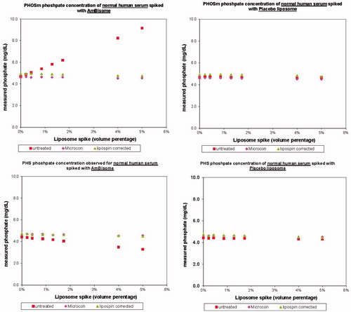
Figure 5. Differential scanning calorimetry scan of AmBisome liposomes.
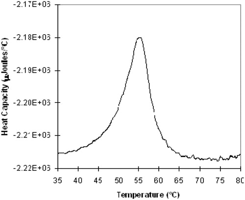
Figure 6. Summary of chemical and physical stability of AmBisome as lyocake or liquid.
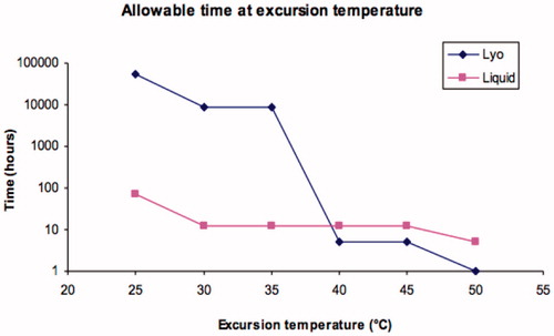
Figure 7. Left: the Temptime instant threshold indicator dot before and after exposure to 40 °C; right: typical application to a vial.
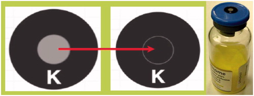
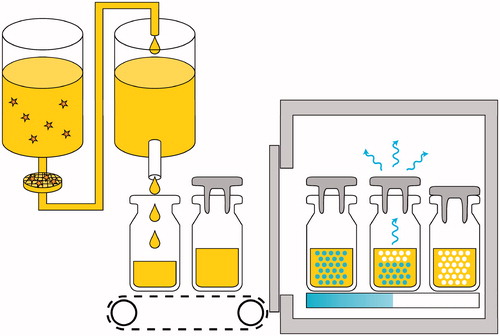
Figure 8. Graphic depiction of bulk formulation, 0.2 micron filtration, fill of sterile bulk, and freeze dry of the liposomal dispersion.