Figures & data
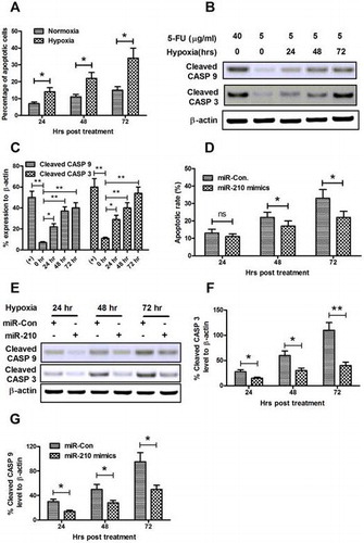
Fig. 1. microRNA-210 is upregulated in bone tissues post-fracture and is positively correlated with HIF-1α levels.
Notes: (A) Relative microRNA-210 expression, examined by RT-qPCR, in normal bone tissues (n = 18) or bone tissues post-fracture (n = 34). (B) Relative HIF-1α mRNA level in bone tissues post-fracture (by RT-qPCR). (C) Correlation between the relative microRNA-210 expressions with the HIF-1α mRNA expression. And statistical significance was considered with a p value <0.05 or less.
Fig. 2. Hypoxia upregulates microRNA-210 expression in osteoblast cells in a HIF-1α-dependent way.
Notes: (A) microRNA-210 expression was upregulated by hypoxia (by RT-qPCR) in osteoblast MG-63 cells. (B) and (C) HIF-1α expression was upregulated by hypoxia at both mRNA (by RT-qPCR) and protein levels (by Western blot assay) in MG-63 cells. (D) Knockdown of HIF-1α by siRNAs transfection for 24 h in MG-63 cells. (E) Abrogation of hypoxia-induced microRNA-210 upregulation by the HIF-1α knockdown (transfection of siHIF-1α 1, siHIF-1α 2, or siRNA control for 24 h). siHIF-1α 1: siRNA-1 targeting siHIF-1α, siHIF-1α 2: siRNA-2 targeting siHIF-1α, siCon: Control siRNA. All experiments were performed in triplicate. Statistical significance was shown as *p < 0.05 and **p < 0.01, ns: no significance.
Fig. 3. Overexpression of microRNA-210 promotes the cellular proliferation of MG-63 cells in vitro.
Notes: (A) The upregulation of microRNA-210 level in MG-63 cells by microRNA-210 mimics transfection. (B) Dose-dependent promotion to the relative cellular proliferation of MG-63 cells in the presence of 5 μg/mL 5-FU by microRNA-210 mimics. (C) Time-dependent promotion to the relative cellular proliferation of MG-63 cells in the presence of 5 μg/mL 5-FU by microRNA-210 mimics. The experiments were performed respectively in triplicate. Statistical significance was shown as *p < 0.05, **p < 0.01, and ***p < 0.001, ns: no significance.
Fig. 4. microRNA-210 mimics stimulated the colony formation of MG-63 cells in vitro.
Notes: Colony formation by MG-63 cells was determined after cells’ transfection with 0 or 50 nM of microRNA-210 mimics or miR-control. The morphologic characteristics were shown of clones formated by MG-63 cells (A); the number of clones formed by MG-63 cells (B) was calculated as comparison. The experiments were performed respectively in triplicate. Statistical significance was shown as ns: no significance, *p < 0.05.
Fig. 5. microRNA-210 mimics ameliorates the hypoxia-induced apoptosis in MG-63 cells.
Notes: (A) Hypoxia-induced apoptosis in MG-63 cells. Cells were treated with 5 μg/mL 5-FU under normoxia or hypoxia condition for 24, 48, or 72 h, then the apoptotic cells were counted by a FACScan flow cytometer. (B) Western blot assay of cleaved CASP 9 and cleaved CASP 3 expression induced by hypoxia. (C) Relative cleaved CASP 9 or cleaved CASP 3 expression as percentage to β-actin. (D) microRNA-210 mimics transfection reduced the hypoxia-induced apoptotic cells. (E–G) microRNA-210 mimics transfection inhibited activated CASP 3 and CASP 9 in MG-63 cells. Cells were transfected with 50 nM microRNA-210 mimics, and were treated with 5 μg/mL 5-FU, under hypoxia condition for 24, 48, or 72 h, then the apoptotic cells were counted by a FACScan flow cytometer; the levels of cleaved CASP 3 and cleaved CASP 9 were determined by Western blot assay. All results were the average of triple experiments. Statistical significance was shown as ns: no significance, *p < 0.05, **p < 0.01, ns: no significance.