Figures & data
Fig. 1. Co-treatment with CP and the VC derivative additively thickened the epidermal and dermal layers in the hairless Sod1−/− mice.
Notes: (A) Hematoxylin and eosin staining of the back skin of hairless Sod1−/− and Sod1+/+ mice treated with the casein peptide (casein), CP (collagen), casein peptide plus the VC derivative (VC) and CP plus the VC derivative. Casein peptide or CP was orally administered, while the VC derivative was transdermally applied on the skin once a day for eight weeks. E, epidermis; D, dermis. The scale bar represents 50 μm (top) and 200 μm (bottom). The thickness of the epidermis (B) and dermis (C) of the back skin of the hairless Sod1−/− mice. The thickness of the epidermis (D) and dermis (E) of the back skin of the hairless Sod1+/+ mice. The data indicate the mean ± SD; *p < 0.05.
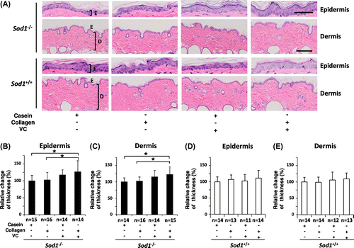
Fig. 2. Treatment with CP and the VC derivative significantly decreased lipid peroxidation in the hairless Sod1−/− skin.
Notes: The (A) MDA and (B) 8-isoprostane content in skin lysates obtained from hairless Sod1−/− male mice treated with the casein peptide only, CP only, casein peptide plus the VC derivative and CP plus the VC derivative. The data indicate the mean ± SD; *p < 0.05, **p < 0.01.
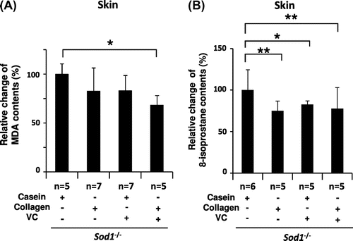
Fig. 3. Treatment with CP and the VC Derivative partially corrected the transcriptional profiles of skin-related genes in the skin tissues of the hairless Sod1−/− mice.
Notes: The relative mRNA expression levels of (A) Col1a1, (B) Mmp2, (C) Has2 and (D) Ki67 were determined using qRT-PCR. The data indicate the mean ± SD; *p < 0.05.
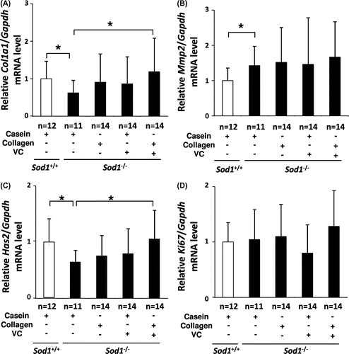
Fig. 4. Treatment with CP and the VC derivative partially normalized the transcriptional profiles of POT family genes in the skin tissues of the hairless Sod1−/− mice.
Notes: The relative mRNA expression levels of (A) Pept1, (B) Pept2, (C) Pht1, and (D) Ci1 were determined using qRT-PCR. The data indicate the mean ± SD; *p < 0.05.
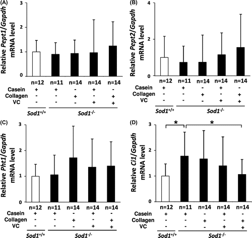
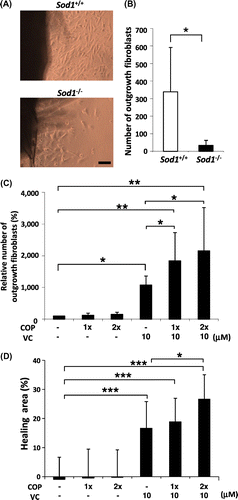
Fig. 5. COP mixture and the VC derivative cooperatively attenuated the impairment of the migration and proliferation of Sod1−/− fibroblasts in vitro.
Notes: (A and B) Number of outgrowth fibroblasts of Sod1+/+ and Sod1−/− mice in the organ culture. (C) Treatment with the COP mixture plus the VC derivative cooperatively promoted the outgrowth activity of the skin fibroblasts. (D) The scratch wound assay allowed for the direct evaluation of migration in vitro. (D) Fibroblasts of Sod1−/− mice were scratched with a pipet tip and cultured for 12 h. (D) Following incubation, the hearing area was quantified by detecting calcein-labeled fibroblasts in the scratch area. 1x and 2x COP indicate one- or twofold concentrations of the COP mixture, respectively. The data indicate the mean ± SD; *p < 0.05, **p < 0.01, ***p < 0.001. The scale bars represent 100 μm.