Figures & data
Table 1. Susceptibility of different indicator strains to nukacin ISK-1 and nisin A.
Fig. 1. Antibacterial activity of nukacin ISK-1.
CFU values (A) immediately after peptide addition (white bars) and 24 h after peptide addition (black bars) in S. simulans and M. luteus are shown. Membrane depolarization (B), ATP release (C), and K+ leakage (nisin is considered 100% release) (D) were measured on S. simulans, M. luteus, and L. sakei cells with no peptide (gray bars), nukacin ISK-1 (white bars), and nisin (black bars) treatment. About 5X MIC of peptides were used for all experiments. The values shown are the means ± standard errors (error bars) for the three independent experiments.
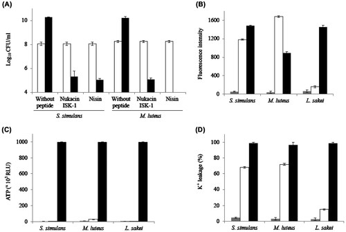
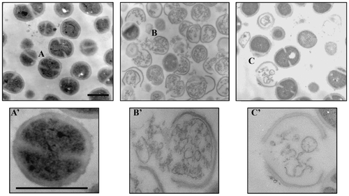
Fig. 2. Transmission electron micrographs of M. luteus cells.
Micrographs of the cells treated without peptide (A), with 10X MIC of nisin (B), and nukacin ISK-1 (C) for 2 h. Enlarged cross-sections of the cells without peptide (A’), with nisin (B’), and nukacin ISK-1 (C’). Bars in panels A and A’: 500 nm.