611
Views
7
CrossRef citations to date
0
Altmetric
Food & Nutrition Science (Notes)

Identification of molecular target of diallyl trisulfide in leukemic cells

, , , , , , , , & show all
Pages 1415-1417 | Received 12 Feb 2014, Accepted 17 Mar 2014, Published online: 12 Jun 2014

Figures & data

Fig. 1. The effect of DATS on cell growth, cell cycle and activation of the proteins regulating apoptosis in U937.

Notes: U937 cells were seeded at a cellular density of 2 × 105 cells/mL and cultured for 24 h. Then the cells were incubated with various concentrations of DATS for 24 h. (A) The cell number was counted by hemocytometer and expressed as a percentage to the vehicle-treated control. (B) Cell cycle distribution analyzed by a flow cytometer. (C) Cleavage of caspase-3 and PARP detected by Western blotting.

Fig. 1. The effect of DATS on cell growth, cell cycle and activation of the proteins regulating apoptosis in U937.Notes: U937 cells were seeded at a cellular density of 2 × 105 cells/mL and cultured for 24 h. Then the cells were incubated with various concentrations of DATS for 24 h. (A) The cell number was counted by hemocytometer and expressed as a percentage to the vehicle-treated control. (B) Cell cycle distribution analyzed by a flow cytometer. (C) Cleavage of caspase-3 and PARP detected by Western blotting.

Fig. 2. Redox 2D-PAGE analysis of U937 treated with DATS.

Notes: (A) Schematic outline of the redox 2D-PAGE. (B) Typical redox 2D-PAGE analysis of U937 treated with DATS. U937 cells were seeded at a cellular density of 2 × 105 cells/mL and cultured for 24 h. Then the cells were treated with 0.2% DMSO as vehicle control (Left panel) or with 10 μM DATS (Right panel) for 10 min and the cell lysates were subjected to the redox 2D-PAGE. The specific spot at approximately 27 kDa was identified to be HSP27.

Fig. 2. Redox 2D-PAGE analysis of U937 treated with DATS.Notes: (A) Schematic outline of the redox 2D-PAGE. (B) Typical redox 2D-PAGE analysis of U937 treated with DATS. U937 cells were seeded at a cellular density of 2 × 105 cells/mL and cultured for 24 h. Then the cells were treated with 0.2% DMSO as vehicle control (Left panel) or with 10 μM DATS (Right panel) for 10 min and the cell lysates were subjected to the redox 2D-PAGE. The specific spot at approximately 27 kDa was identified to be HSP27.

Fig. 3. Formation of HSP27 dimer and cleaved caspase-3 in DATS-treated U937.

Notes: U937 cells were treated with 10 μM DATS for the times indicated in the panels. (A) HSP27 dimer formation was evaluated by non-reduced SDS-PAGE followed by Western blotting. (B) HSP27 dimer formation and the cleavage of caspase-3 were detected by Western blotting. U937 cells were also pretreated with GSH (1 mM) for 30 min prior to the DATS treatment (DATS + GSH).
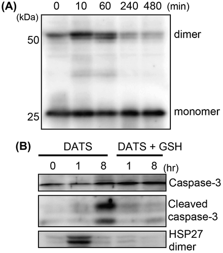
Fig. 3. Formation of HSP27 dimer and cleaved caspase-3 in DATS-treated U937.Notes: U937 cells were treated with 10 μM DATS for the times indicated in the panels. (A) HSP27 dimer formation was evaluated by non-reduced SDS-PAGE followed by Western blotting. (B) HSP27 dimer formation and the cleavage of caspase-3 were detected by Western blotting. U937 cells were also pretreated with GSH (1 mM) for 30 min prior to the DATS treatment (DATS + GSH).

Reprints and Corporate Permissions

Please note: Selecting permissions does not provide access to the full text of the article, please see our help page How do I view content?

To request a reprint or corporate permissions for this article, please click on the relevant link below:

Academic Permissions

Please note: Selecting permissions does not provide access to the full text of the article, please see our help page How do I view content?

Obtain permissions instantly via Rightslink by clicking on the button below:

If you are unable to obtain permissions via Rightslink, please complete and submit this Permissions form. For more information, please visit our Permissions help page.