Figures & data
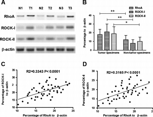
Fig. 1. Overexpression of RhoA and ROCK-I/II in CC specimens.
Notes: (A) and (B) overexpression of RhoA and ROCK-I/II in part CC specimens; and (C) and (D) correlation of RhoA overexpression with the ROCK-I(C) or ROCK-II (D) expression. Statistical significance was considered with a p value < 0.05 or less.
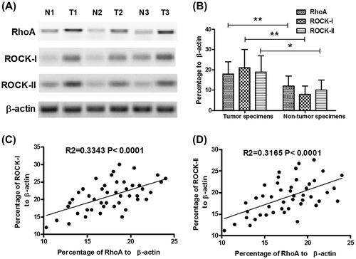
Fig. 2. Association of RhoA overexpression with the clinicopathological variants of CCs.
Notes: (A) RhoA expression of CC specimens from patients with different FIGO stages; (B) RhoA expression of CC specimens from patients with or without vascular invasion; and (C) RhoA expression of CC specimens from patients with or without lymph node metastasis. Statistical significance was considered with a p value < 0.05 or less.
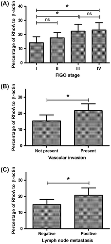
Fig. 3. Construction of a Hela cell line, overexpressing RhoA.
Notes: (A) schematic diagram of RhoA and EGFP co-expression with a 2A peptide sequence; the RhoA and EGFP coding sequence linked by 2A peptide coding sequence was cloned into pcDNA3.1 vector; a single mRNA coding CTGF and RFP could translate RhoA and EGFP separately; (B) EGFP expression of Hela (RhoA+) cells; (C) RhoA expression in protein level in the Hela (RhoA+) cells; (D) RhoA expression in mRNA level in the Hela (RhoA+) cells; and (E) and (F) knockdown of RhoA in mRNA level (E) or in protein level (F) by siRNAs. All results are the average of three independent experiments, and statistical significance was considered with a p value < 0.05 or less.
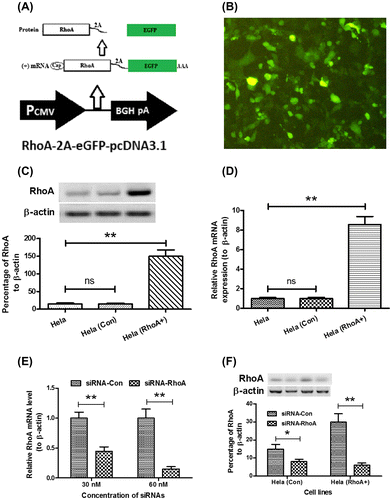
Fig. 4. Rho promotes CC cell proliferation.
Notes: (A) significantly higher proliferation of Hela (RhoA+) cells than Hela (Con) cells. The growth curve of both cells was evaluated by CCK-8 assay. (B) growth curve of Hela (RhoA+) cells after transfecting with siRNA-RhoA or siRNA-Con, examined by CCK-8 assay. The experiments were performed separately in triplicate. Statistical significance was shown as *p < 0.05, **p < 0.01.
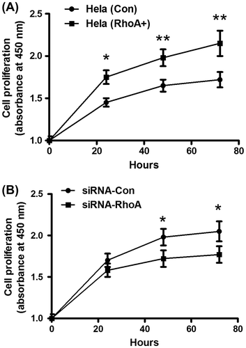
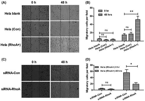
Fig. 5. Rho promotes CC cell migration.
Notes: (A) and (B) higher migration of Hela, Hela (RhoA+) cells than Hela (Con) cells. Migration of cells was depicted post inoculation at 0, or 48 h by cells scratch assay. Solid lines showed the edges at the start of experiments. (C) and (D) inhibition by siRNA-RhoA, of the promotion of RhoA to the Hela cell migration. Migration of Hela (RhoA+) cells was depicted post transfecting with siRNA-RhoA or siRNA-Con at 0, or 48 h by cells scratch assay. The experiments were performed separately in triplicate. Statistical significant was showed as *p < 0.05 and **p < 0.01, ns: not significant.