Figures & data

Table 1. Diet compositions.
Table 2. Effects of biotin deficiency.
Table 3. Effects of biotin restoration.
Fig. 1. Spindle abnormalities in oocytes induced by biotin deficiency.
Notes: Representative examples of meiotic spindles in oocytes from indicated mice (n = 37–38 oocytes analyzed per group) after labeling α-tubulin antibody (green) and counter-staining DNA with DAPI (aqua blue). (A) control group, (B) biotin-deficient group.
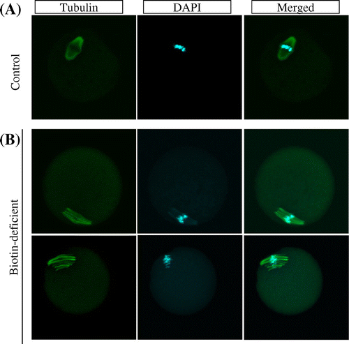
Fig. 2. Higher frequency of abnormal oocytes induced by biotin deficiency not restored by re-feeding of biotin-containing diet.
Notes: Abnormal oocyte number is the summation of chromosome alignment and spindle defect in oocytes. (A) Day 22, (B) Day 42. (C) control; BD; biotin deficient group; R, recovery group. n = 37–54 oocytes analyzed per group.
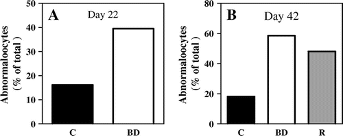
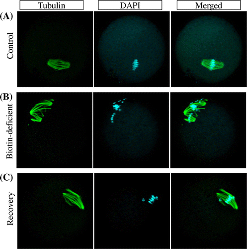
Fig. 3. Meiosis aneuploidy induced by biotin deficiency, not restored by biotin re-feeding.
Notes: Representative examples of meiotic spindles in oocytes from indicated mice (n = 41–66 oocytes analyzed per group) after labeling α-tubulin antibody (green) and counter-staining DNA with DAPI (aqua blue). (A) control group, (B) biotin-deficient group, and, (C) recovery group.