
Figures & data
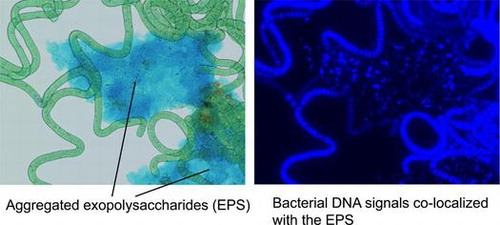
Fig. 1. Contaminating bacteria in subfractions of non-axenic cultures.
Notes: (A) Growth of contaminating bacteria. (B) Contaminating bacteria in the aqueous part of the suspension of washed trichomes. (C) Contaminating bacteria associated with trichomes. To determine growth of contaminating bacteria, trichomes were removed from a non-axenic culture (1 mL) of A. platensis UTEX 1926 (1.8 × 10³ trichomes/mL) by filtering through a track-etched polycarbonate membrane with 8-μm openings. This filtrate containing free-floating bacteria was incubated at 30 °C. At the indicated time points, samples were withdrawn, and cfus of heterotrophic bacteria were determined. The data in (A) represent the averages of five measurements, and error bars indicate standard errors of the means. In the experiment in (B), to determine the cfu of contaminating bacteria in the aqueous part of the suspension of washed trichomes, a non-axenic culture (1 mL) was applied on a nylon mesh with 20-μm openings, and the trichomes on the mesh were washed with 50 mL of sterile SOT medium. Washed trichomes were then suspended in 1 mL of the same medium and incubated at 30 °C. At the indicated time points, 5 μL aliquots of the medium were withdrawn with a micropipette so as to not contain trichomes, and cfu were determined. The data in (B) represent the averages of five independent filtration experiments beginning with a single non-axenic culture, and error bars indicate the standard errors of the means. In the experiment in (C), to detect contaminating bacteria associated with trichomes, at the indicated time points, six trichomes with 1 μL each of medium were withdrawn using a micropipette from the trichome suspension prepared in the experiment in (B), and each of these was individually washed by serial transfer to three droplets (0.3 mL each) of sterile SOT medium. After washing, trichomes in 1 μL of the medium were placed on a solidified SOT medium supplemented with yeast extract. Trichomes on the agar plates were incubated at 30 °C for 72 h with occasional observation under a dissecting microscope to detect the growth of any contaminating bacteria around the trichomes.
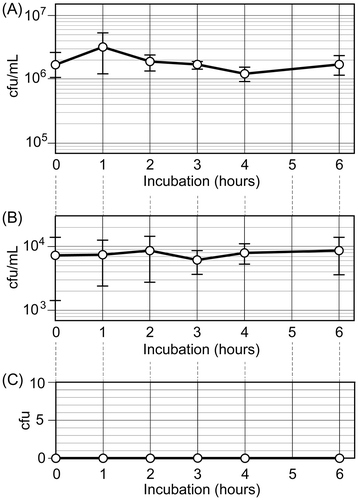
Fig. 2. Alcian blue staining for the preparations of Arthrospira trichomes.
Notes: (A) Bright-field view of a trichome preparation from a non-axenic culture of A. platensis UTEX 1926. (B) Alcian blue-stained non-axenic culture of A. platensis UTEX 1926. (C) Alcian blue-stained axenic culture of A. platensis NIES-39. (D) Alcian blue-stained axenic culture of A. platensis NIES-46. Scale bars represent 100 μm.
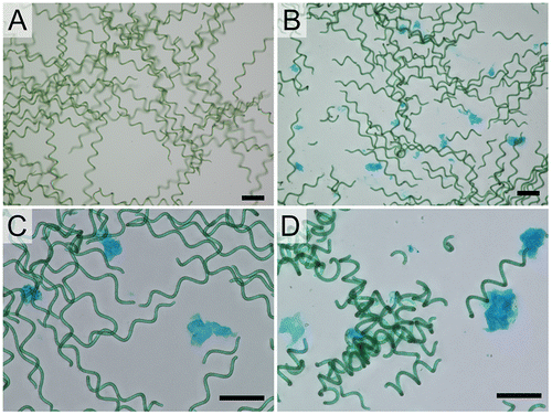
Fig. 3. Association of bacteria with aggregates of EPS in non-axenic cultures.
Notes: Preparations of trichomes from non-axenic and axenic cultures of A. platensis UTEX 1926 were double-stained with Alcian blue and Hoechst 33342. (A–C) Preparation from a non-axenic culture. (D–F) Preparation from an axenic culture. (A) and (D) Bright-field views. (B) and (E) Fluorescence images. (C) and (F) Chlorophyll autofluorescence. Scale bars represent 20 μm.
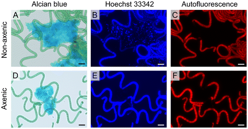
Table 1. Isolation of axenic trichomes of various Arthrospira strains.
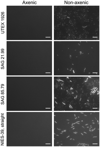
Fig. 4. Heterotrophic bacteria in the cultures prepared in this study and in original non-axenic cultures.
Notes: Cultures of A. platensis UTEX 1926, A. platensis SAG 21.99, A. platensis SAG 85.79, and a straight variant of A. platensis NIES-39 were filtered through track-etched membranes with 8-μm pores to eliminate Arthrospira trichomes. Heterotrophic bacteria in the filtrates were collected on track-etched membranes with 0.2-μm pores and stained with DAPI. Materials from 5 mL of cultures were collected for the preparations obtained in this study (Axenic), whereas those from 0. 2 mL were collected from non-axenic cultures (Non-axenic). Scale bars represent 5 μm.
Supplemental material