763
Views
17
CrossRef citations to date
0
Altmetric
Environmental Science

A mechanism for nano-titanium dioxide-induced cytotoxicity in HaCaT cells under UVA irradiation

, &
Pages 1384-1390 | Received 24 Dec 2014, Accepted 06 Feb 2015, Published online: 30 Mar 2015

Figures & data

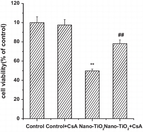
Fig. 1. Effects of MPT pore in nano-TiO2-induced cell viability and ROS formation of HaCaT cells.

Notes: Effects of MPT pore in nano-TiO2 induced-(B) cell viability and (C) ROS formation of HaCaT cells. Cells were treated with 200 μg/mL nano-TiO2 only, or pretreated with CsA (10.0 μM) for 30 min, followed by treatment with 200 μg/mL nano-TiO2. Control was received culture medium only. All samples were irradiated with the UVA light for 1 h and then cultured for 24 h. Results are expressed as mean ± SEM of at least four different experiments (**p < 0.01 represents the comparison with the control group; ##p < 0.01 represents the comparison with the nano-TiO2 group).
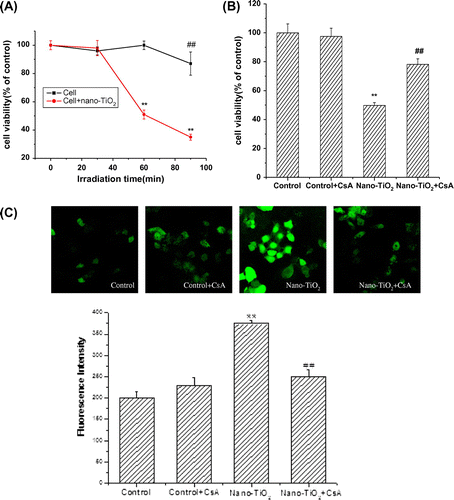
Fig. 1. Effects of MPT pore in nano-TiO2-induced cell viability and ROS formation of HaCaT cells.Notes: Effects of MPT pore in nano-TiO2 induced-(B) cell viability and (C) ROS formation of HaCaT cells. Cells were treated with 200 μg/mL nano-TiO2 only, or pretreated with CsA (10.0 μM) for 30 min, followed by treatment with 200 μg/mL nano-TiO2. Control was received culture medium only. All samples were irradiated with the UVA light for 1 h and then cultured for 24 h. Results are expressed as mean ± SEM of at least four different experiments (**p < 0.01 represents the comparison with the control group; ##p < 0.01 represents the comparison with the nano-TiO2 group).
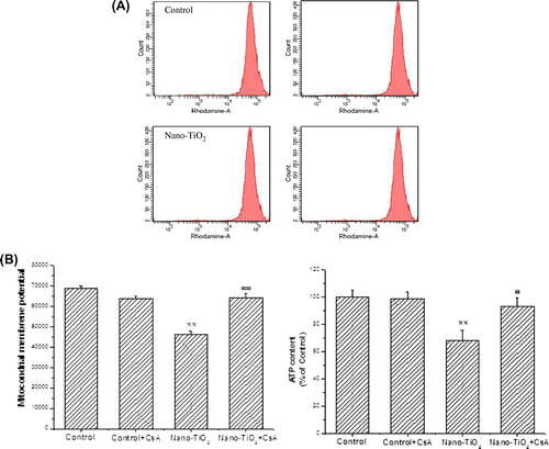
Fig. 2. Effects of MPT pore on MMP decrease and ATP depletion of HaCaT cells.

Notes: Effects of MPT pore on (A) MMP decrease and (B) ATP depletion of HaCaT cells. Cells were treated with 200 μg/mL nano-TiO2 only, or pretreated with CSA (10 μM) for 30 min, followed by treatment with 200 μg/mL nano-TiO2. Control was received culture medium only. All samples were irradiated with the UVA light for 1 h and then cultured for 24 h. Results are expressed as mean ± SEM. of at least four different experiments (**p < 0.01 represents the comparison with the control group; ##p < 0.01 represents the comparison with the nano-TiO2 group).
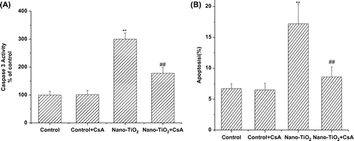
Fig. 2. Effects of MPT pore on MMP decrease and ATP depletion of HaCaT cells.Notes: Effects of MPT pore on (A) MMP decrease and (B) ATP depletion of HaCaT cells. Cells were treated with 200 μg/mL nano-TiO2 only, or pretreated with CSA (10 μM) for 30 min, followed by treatment with 200 μg/mL nano-TiO2. Control was received culture medium only. All samples were irradiated with the UVA light for 1 h and then cultured for 24 h. Results are expressed as mean ± SEM. of at least four different experiments (**p < 0.01 represents the comparison with the control group; ##p < 0.01 represents the comparison with the nano-TiO2 group).

Fig. 3. Effects of MPT pore in nano-TiO2-induced Caspase-3 activation and cell apoptosis of HaCaT cells.

Notes: Effects of MPT pore in nano-TiO2-induced (A) Caspase-3 activation and (B) cell apoptosis of HaCaT cells. Cells were treated with 200 μg/mL nano-TiO2 only, or pretreated with CsA (10.0 μM) for 30 min, followed by treatment with 200 μg/mL nano-TiO2. Control was received culture medium only. All samples were irradiated with the UVA light for 1 h and then cultured for 24 h. Results are expressed as mean ± SEM of at least four different experiments (**p < 0.01 represents the comparison with the control group; ##p < 0.01 represents the comparison with the nano-TiO2 group).

Fig. 3. Effects of MPT pore in nano-TiO2-induced Caspase-3 activation and cell apoptosis of HaCaT cells.Notes: Effects of MPT pore in nano-TiO2-induced (A) Caspase-3 activation and (B) cell apoptosis of HaCaT cells. Cells were treated with 200 μg/mL nano-TiO2 only, or pretreated with CsA (10.0 μM) for 30 min, followed by treatment with 200 μg/mL nano-TiO2. Control was received culture medium only. All samples were irradiated with the UVA light for 1 h and then cultured for 24 h. Results are expressed as mean ± SEM of at least four different experiments (**p < 0.01 represents the comparison with the control group; ##p < 0.01 represents the comparison with the nano-TiO2 group).

Fig. 4. Effects of MPT pore in nano-TiO2-induced K6 mRNA expression.

Notes: Effects of MPT pore in nano-TiO2-induced K6 mRNA expression. Cells were treated with 200 μg/mL nano-TiO2 only, or pretreated with CsA (10.0 μM) for 30 min, followed by treatment with 200 μg/mL nano-TiO2. Control was received culture medium only. All samples were irradiated with the UVA light for 1 h and then cultured for 24 h. Results are expressed as mean ± SEM of at least four different experiments (*p < 0.05 represents the comparison with the control group; #p < 0.05 represents the comparison with the nano-TiO2 group).
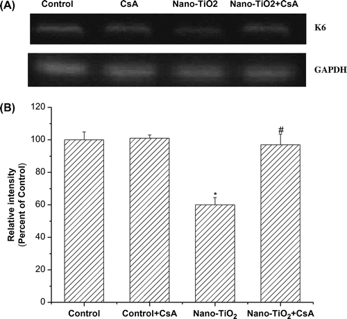
Fig. 4. Effects of MPT pore in nano-TiO2-induced K6 mRNA expression.Notes: Effects of MPT pore in nano-TiO2-induced K6 mRNA expression. Cells were treated with 200 μg/mL nano-TiO2 only, or pretreated with CsA (10.0 μM) for 30 min, followed by treatment with 200 μg/mL nano-TiO2. Control was received culture medium only. All samples were irradiated with the UVA light for 1 h and then cultured for 24 h. Results are expressed as mean ± SEM of at least four different experiments (*p < 0.05 represents the comparison with the control group; #p < 0.05 represents the comparison with the nano-TiO2 group).

Reprints and Corporate Permissions

Please note: Selecting permissions does not provide access to the full text of the article, please see our help page How do I view content?

To request a reprint or corporate permissions for this article, please click on the relevant link below:

Academic Permissions

Please note: Selecting permissions does not provide access to the full text of the article, please see our help page How do I view content?

Obtain permissions instantly via Rightslink by clicking on the button below:

If you are unable to obtain permissions via Rightslink, please complete and submit this Permissions form. For more information, please visit our Permissions help page.