Figures & data
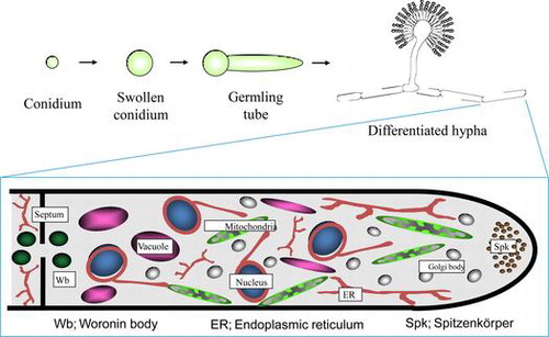
Fig. 1. Schematic drawing of the A. oryzae genome.Citation4)
Fig. 2. Visualization of organelles in A. oryzae by expression of EGFP fusion proteins. (A) Endoplasmic reticulum,Citation7) (B) Golgi apparatus,Citation7) (C) mitochondria,Citation9) (D) vacuoles,Citation10) (E), peroxisomes,Citation11) and (F), nuclei.
Fig. 3. Schematic model of Aspergillus oryzae organelles.
Fig. 4. Localization of α-amylase fused with EGFP in liquid and agar cultures.Citation16).
Fig. 5. Proposed model for the endocytic recycling in apical tip supporting high protein secretion of A. oryzae.Citation22)
Fig. 6. Pleiomorphic vacuoles in A. oryzae.Citation23)
Fig. 7. Schematic diagram for the putative flow of nutrients recycled by autophagy during conidiation and germination.Citation27)
Fig. 8. Dual fluorescent images of an A. oryzae strain expressing DsRed2–AoHex1 and RNase T1–EGFP fusion proteins.Citation15)