Figures & data
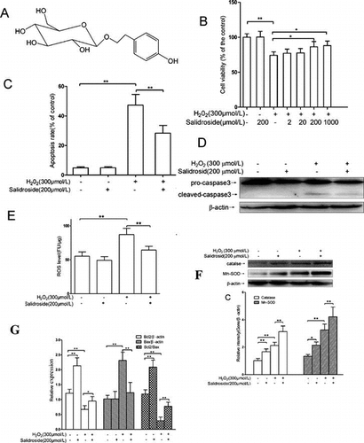
Fig. 1. Chemical structure of salidroside.
Table 1. Sequences of the primers used for real-time PCR amplification.
Fig. 2. Salidroside protected primary-cultured RECs against H2O2-induced apoptosis.
Notes: (A) After incubation with H2O2 (0–700 μmol/L) for 4 h, REC viability was measured by WST-1 assay. (B) Effects of salidroside on cell viability measured by WST-1 assay (n = 5/group). (C) Intracellular apoptotic nuclei detected by TUNEL assay. Yellow arrows indicate the purplish red-stained TUNEL-positive nuclei. Scale bars = 50 μm, Original magnifications ×400 (n = 5/group). (D) Quantitative analysis of RECs apoptosis. (E) Pro- and cleaved caspase 3 proteins detected by Western blotting. RECs were treated by salidroside alone for 12 h, or H2O2 alone for 4 h, or salidroside pre-incubation for 12 h and then co-incubation with H2O2 for another 4 h. (F) Densitometric analysis of cleaved caspase-3 (n = 5/group). *p < 0.05, **p < 0.01.
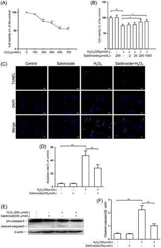
Fig. 3. Effects of salidroside on oxidative stress markers, MDA, and ROS in primary-cultured RECs.
Notes: (A) MDA contents in retinal endothelial cells (n = 5/group). (B) Intracellular ROS was measured using H2DCFDA and normalized to the protein contents in cell lysate (n = 6/group). **p < 0.01.
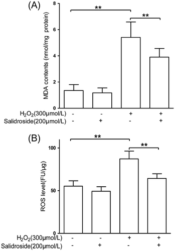
Fig. 4. Activities and expression of catalase and Mn-SOD in primary-cultured RECs.
Notes: mRNA expression (A), protein levels (B and C), and activities (D) of catalase and Mn-SOD in RECs treated by either salidroside alone or H2O2 with or without salidroside (n = 4/group). (*p < 0.05, **p < 0.01).
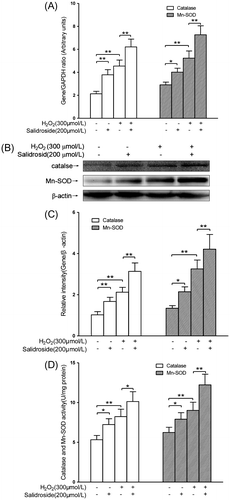
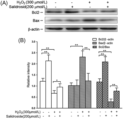
Fig. 5. Effects of salidroside on the expressions of Bcl2 and Bax in primary-cultured RECs.
Notes: (A) Intracellular Bcl2/Bax expressions detected by Western blot analysis. (B) The quantitative comparisons of Bcl2, Bax, and Bcl2/Bax (n = 4/group). *p < 0.05, **p < 0.01.