
Figures & data
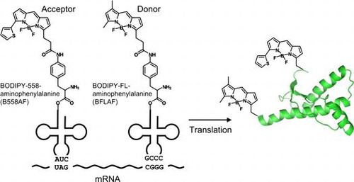
Fig. 1. Schema of double-fluorescent labeled PrP synthesis.
Notes: B558AF and BFLAF were incorporated into PrP using amber and four-base codons.
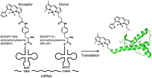
Fig. 2. The introduction of mutations into expression vector.
Notes: Boldfaced amino acids show the amino acid sequence of mPrP(121–231). TAG (amber) and CGGG four-base codons were inserted into N- and C-terminal sites, respectively, of the mPrP(121–231) gene. A stop codon TAA after the amino acid residue of 124 appears when CGGG is decoded as a triplet codon, but the expressed protein is not purified using a His SpinTrap column. TAG amber codon was also substituted for the codon of the mPrP(121–231) gene encoding Tyr127, Phe140, Phe174, Phe197, and Tyr217. The gene sequence of the protein 140/232 after the optimization is shown in the middle line (gray).
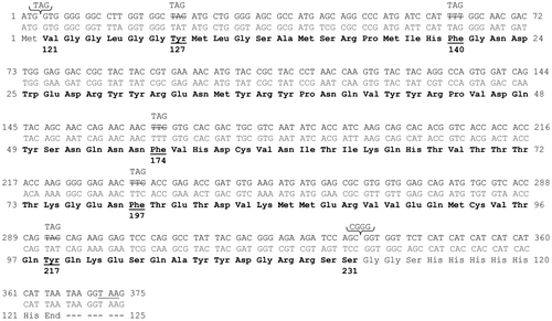
Table 1. Concentrations of double-fluorescent labeled PrP synthesized in this study.
Fig. 3. Analysis of purified, double-labeled protein containing BFLAF and B558AF.
Notes: (A) Fluorescence image of SDS-PAGE. Top: excitation at 488 nm and emission at 520 nm. Middle: excitation at 532 nm and emission at 580 nm. Bottom: merged image of top and middle. (B) Western blot analysis using an antibody against PrP (M-20). FLM: BenchMarkTM Fluorescent Protein Standard (Invitrogen). PSM: Prestained Protein Marker, Broad Range (NEB).
Fig. 4. Each double-labeled protein 140/232 from the original and optimized sequences was separated into total fraction (pre-separation, T), supernatant fraction (S), and pellet fraction (P).
Notes: Aliquots of the reaction mixtures (2 μL) were loaded onto a SDS-PAGE gel. (A) Merged image with excitation at 488 nm and emission at 520 nm, and excitation at 532 nm and emission at 580 nm. (B) Western blot analysis using anti-penta-His antibody. FLM: BenchMarkTM Fluorescent Protein Standard (Invitrogen). M: BenchMarkTM Protein Ladder (Invitrogen).
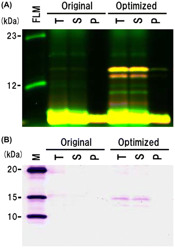
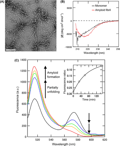
Fig. 5. Amyloid fibril formation of the double-labeled protein 120/232.
Notes: (A) EM image of mPrP(23–231) amyloid fibrils, used as seeds for amyloid formation of the double-labeled PrP. The scale bar indicates 200 nm. (B) CD spectra of monomer (black) and amyloid fibrils (red) of mPrP(23–231) in 50 mM MES-NaOH buffer (pH 5.5) containing 2 M GdnHCl. (C) Fluorescence spectra of the double-labeled protein 120/232 with an excitation at 488 nm. Black line shows the fluorescence spectrum of the protein 120/232 at the native conditions. Blue, green, orange, and red lines show the fluorescence spectra after incubations of 0, 30, 60, and 150 min, respectively, under the ultrasonication. Inset shows the fluorescence ratio at 573 and 515 nm (acceptor and donor, respectively) with incubation time.
Supplemental material