Figures & data
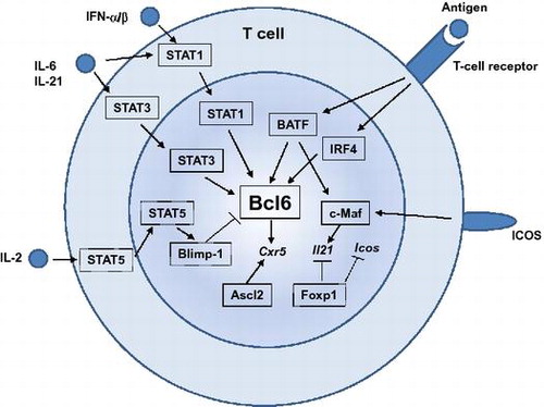
Fig. 1. Differentiation of naïve CD4+T cells into distinct helper T-cell subsets.
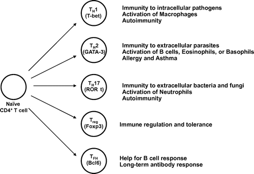
Fig. 2. Multiple steps involved in TFH-cell development.
Notes: TFH-cell development requires serial interaction with DCs and antigen-specific B cells. Upon priming with dendritic cells, some of the activated T cells up-regulate a chemokine receptor, CXCR5, and transcription factors, including Bcl6 (a). The activated CD4+ T cells migrate toward the border of T-cell zone and B-cell follicle, where the T cells interact with antigen-specific B cells (b). This interaction with B cells further up-regulates CXCR5 and Bcl6, while down-regulates CCR7, which allows the T cells relocate to B-cell follicle (TFH) (c). Some of TFH cells participate in germinal center (GC) reaction (GC-TFH) (d).
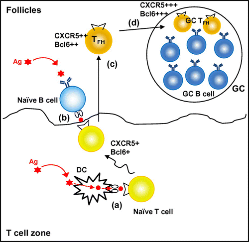
Fig. 3. Transcription factors that regulate TFH-cell development.
Notes: Many transcription factors are involved in TFH-cell development. Transcription factors shown in boxes with straight lines are the positive regulators, whereas those in boxes with dashed lines are the negative regulators for TFH development. Among them, Bcl6 plays a central role during TFH-cell development. STAT1, STAT3, BATF, and IRF4 positively regulate Bcl6 induction, whereas a STAT5-Blimp1 axis inhibits Bcl6 induction. Ascl2 is involved in CXCR5 expression. c-Maf is required for optimal IL-21 expression. Foxp1 negatively regulates IL-21 or ICOS expression.
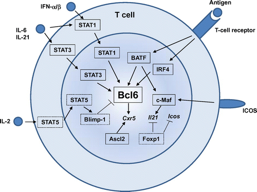
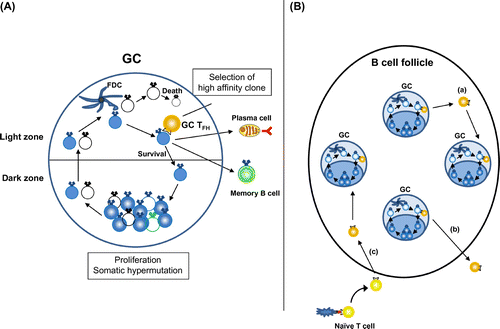
Fig. 4. Function of TFH cells in GCs.
Notes: (A) GC-TFH cells localize in the light zone of GCs and scan the amount of antigenic peptide presented on GC B cells. GC B cells with high-affinity BCRs present high levels of antigenic peptide and get survival signals from GC-TFH cells. Selected high-affinity B cells differentiate into plasma cells or memory B cells, or migrate back to the dark zone of GC to undertake further round of proliferation and somatic hypermutation. (B) GC-TFH cells are not confined to GC. GC-TFH cells can emigrate to the follicles and enter different GCs (a) or relocate to the T-cell zone (b). Further, newly generated TFH cells can participate in preexisting GCs (c).