Figures & data
Fig. 1. (A) The structure and transmission electron microscopy (TEM) image of a small glass particle incorporating multiple CdSe/ZnS QDs (prepared at AIST) and (B) The structure of a CdSe/ZnS QD coated with polymer (commercially available).
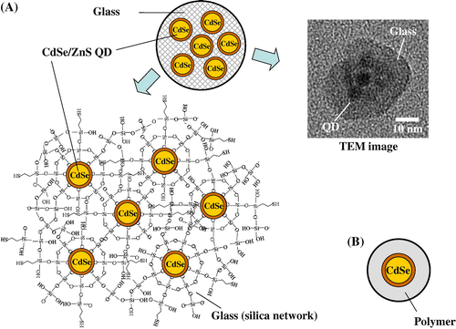
Table 1. Nanoparticle samples used for testing their cytotoxicity.
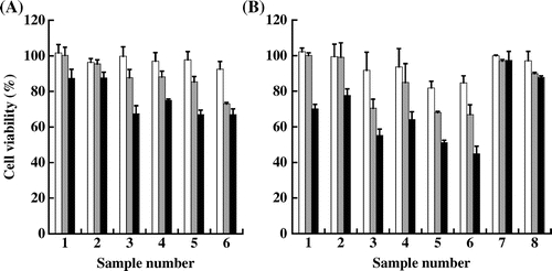
Fig. 2. Cell viability of HaCaT cells after exposure to the nanoparticle samples listed in Table for 6 h (A) and 24 h (B).
Notes: White, gray and black columns show cell viability after exposure to nanoparticles (NPs) at QD concentration of 0.1, 1.0 and 10 nM, respectively. The values indicate means ± S.D. of triple determinations.