1,234
Views
12
CrossRef citations to date
0
Altmetric
Biochemistry & Molecular Biology

Anti-skeletal muscle atrophy effect of Oenothera odorata root extract via reactive oxygen species-dependent signaling pathways in cellular and mouse model

, , , , , , & show all
Pages 80-88 | Received 03 Apr 2015, Accepted 14 Jul 2015, Published online: 19 Aug 2015

Figures & data

Fig. 1. Effects of intraperitoneal injection of EVP on disuse muscle atrophy by sciatic denervation in mice.
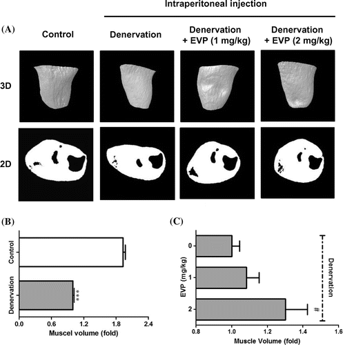
Notes: (A) Muscle volume was measured by in vivo micro-CT. Two-dimensional (2D) cross-sectional images positioned 3.6 mm below the growth plate of tibia and three-dimensional (3D) reconstruction models of muscle are shown. Images of muscle in sciatic-denervated mice (n = 5) were acquired at 21 days after the induced muscle atrophy using the micro-CT. (B and C) For the evaluation of muscle volume, 3D and 2D reconstructed images using CT-Analyzer 1.11 in the control, sciatic denervated, and intraperitoneal injection of EVP (1–2 mg/kg) groups. Each value represents the mean (±SD) from five mice. ***p < 0.0001 vs. control, #p < 0.01 vs. sciatic denervation alone.
Fig. 1. Effects of intraperitoneal injection of EVP on disuse muscle atrophy by sciatic denervation in mice.

Fig. 2. Effects of EVP on substances and proteins relating to the pathway between ROS and apoptosis in muscle of sciatic-denervated mice.
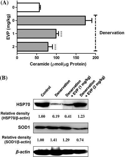
Notes: (A) Effect of EVP on ceramide level by disuse muscle atrophy in mice. Lipids were extracted from whole lysate of muscle with ethanol containing C17-ceramide as internal standard. Ceramide quantification was performed by HPLC. (B) Effect of EVP on HSP70 and SOD1 expression by disuse muscle atrophy in mice. SOD1, HSP70, and β-actin were analyzed by immunoblot analysis using specific antibodies. Each value represents the mean from five mice. ***p < 0.0001 vs. sciatic denervation alone.
Fig. 2. Effects of EVP on substances and proteins relating to the pathway between ROS and apoptosis in muscle of sciatic-denervated mice.

Fig. 3. Effect of EVP on H2O2-induced oxidative stress in C2C12 myoblasts.
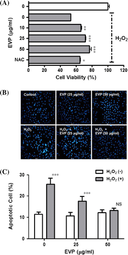
Notes: (A) Effect of EVP on H2O2-induced cell viability in C2C12 myoblasts. The optical density was set at 450 nm using a microplate reader. The cell viability was calculated using the following equation: Cell viability (%) = [(absorbance of the H2O2-treated sample/absorbance of the H2O2-untreated control) × 100]. (B and C) Effect of EVP on apoptosis by oxidative stress in C2C12 myoblasts. The apoptotic cells were estimated by direct counting after staining. Each value represents the mean (±SD) from three experiments, each performed in triplicate. NAC: N-acetyl cysteine, DAPI: 4, 6-diamino-2-phenylindole. NAC: 2 mM. *p < 0.01, **p < 0.001 and ***p < 0.0001 vs. H2O2 alone. NS: Not significant.
Fig. 3. Effect of EVP on H2O2-induced oxidative stress in C2C12 myoblasts.

Fig. 4. Effect of EVP on ceramide level in H2O2-induced oxidative stress in C2C12 myoblasts.
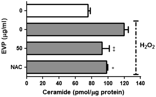
Notes: Lipids were extracted from the C2C12 myoblast pellets with ethanol containing C17-ceramide as an internal standard. Ceramide quantification was performed by HPLC. Each value represents the mean (±SD) from three experiments. NAC: 2 mM. NAC: N-acetyl cysteine. **p < 0.001 vs. H2O2 alone.
Fig. 4. Effect of EVP on ceramide level in H2O2-induced oxidative stress in C2C12 myoblasts.

Fig. 5. Effect of EVP on oxidative stress induced by ROS in C2C12 myoblasts.
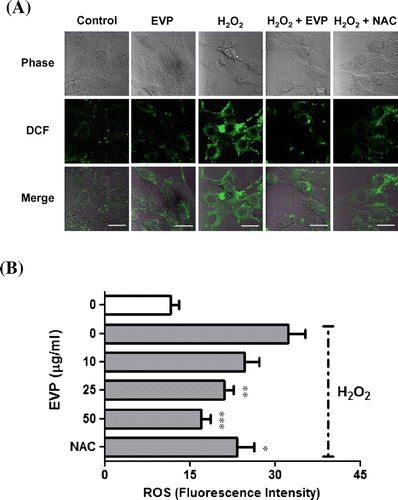
Notes: (A) Effect of EVP on intracellular ROS in C2C12 myoblasts using confocal microscope. Representative pictures of ROS production were acquired with a Zeiss LSM710 confocal microscope. (B) Effect of EVP on intracellular ROS in C2C12 myoblasts using microplate reader. The relative levels of fluorescence were quantified with an FLx800 microplate fluorescence reader at the excitation and emission wavelengths of 485 and 535 nm, respectively. Each value represents the mean (±SD) from three experiments, each performed in triplicate. NAC: N-acetyl cysteine, DCF-DA: 2′,7′-dichlorofluorescein diacetate. NAC: 2 mM. EVP: 50 µg/mL. *p < 0.01, **p < 0.001, and ***p < 0.0001 vs. H2O2 alone.
Fig. 5. Effect of EVP on oxidative stress induced by ROS in C2C12 myoblasts.

Fig. 6. Effect of EVP on SOD1 expression in H2O2-induced oxidative stress in C2C12 myoblasts.
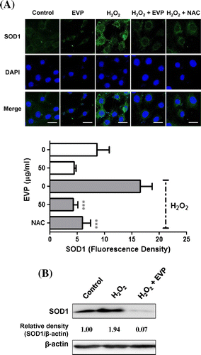
Notes: (A) Effect of EVP on SOD1 expression by ROS in C2C12 myoblasts. Representative images (up) and quantitative analysis (down) of SOD1 (green) are shown for C2C12 myoblasts. C2C12 myoblasts were cultured on a cover glass with DMSO (maximally 0.05%), NAC (2 mM) or EVP (50 µg/mL) prior to pre-incubating for 23 h; H2O2 (1 mM) was added for 1 h. Images were acquired with a LSM710 confocal microscope. Shown are the mean values (±SD) from three experiments. Scale bar: 20 µm. NAC: N-acetyl cysteine. (B) Effect of EVP on SOD1 protein level by oxidative stress in C2C12 myoblasts. SOD1 and β-actin were analyzed by immunoblot analysis using specific antibodies. Each value represents the mean from three experiments. EVP: 50 µg/mL. ***p < 0.0001 vs. H2O2 alone.
Fig. 6. Effect of EVP on SOD1 expression in H2O2-induced oxidative stress in C2C12 myoblasts.

Fig. 7. Effect of EVP on HSP70 expression in H2O2-induced oxidative stress in C2C12 myoblasts.
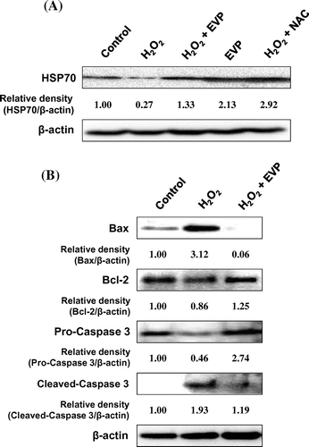
Notes: (A) Effect of EVP on HSP70 protein level by oxidative stress in C2C12 myoblasts. (B) Effect of EVP on expression of pro- to anti-apoptotic protein including Bax, Bcl-2, caspase 3, and β-actin by oxidative stress in C2C12 myoblasts. Each protein was analyzed by immunoblot analysis using specific antibodies. Each value represents the mean from three experiments. NAC: 2 mM. EVP: 50 µg/mL. NAC: N-acetyl cysteine.
Fig. 7. Effect of EVP on HSP70 expression in H2O2-induced oxidative stress in C2C12 myoblasts.

Reprints and Corporate Permissions

Please note: Selecting permissions does not provide access to the full text of the article, please see our help page How do I view content?

To request a reprint or corporate permissions for this article, please click on the relevant link below:

Academic Permissions

Please note: Selecting permissions does not provide access to the full text of the article, please see our help page How do I view content?

Obtain permissions instantly via Rightslink by clicking on the button below:

If you are unable to obtain permissions via Rightslink, please complete and submit this Permissions form. For more information, please visit our Permissions help page.