Figures & data
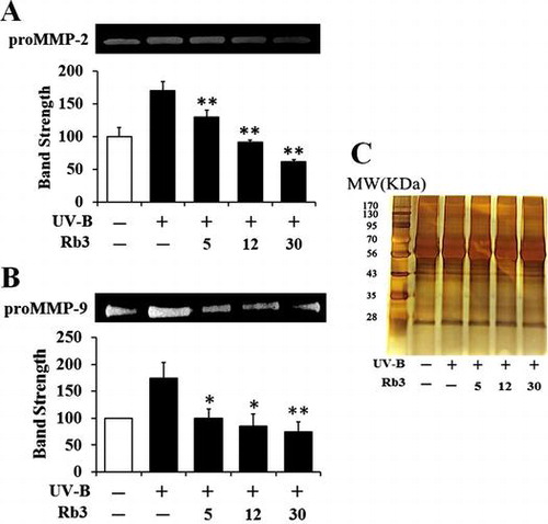
Fig. 1. The chemical structure of ginsenoside Rb3 (Rb3).
Fig. 2. Effect of Rb3 on reactive oxygen species (ROS) elevation in HaCaT keratinocytes under irradiation with 70 mJ/cm2 UV-B radiation. HaCaT cells were subjected to fresh medium with the indicated concentrations (0, 5, 12, or 30 μM) of Rb3 for 30 min prior to the irradiation. The intracellular ROS level was quantitated using DCFH-DA in a microplate fluorometer (A) and dihydrorhodamine 123 in confocal microscopic analysis (B).
Fig. 3. Effect of Rb3 on cellular viability in HaCaT keratinocytes under irradiation with 70 mJ/cm2 UV-B radiation.
Fig. 4. Effect of Rb3 on the gelatinolytic activities of proMMP-2 (A) and proMMP-9 (B) in the conditioned media obtained from HaCaT keratinocyte cultures under irradiation with 70 mJ/cm2 UV-B radiation.
Fig. 5. Effect of Rb3 on proMMP-2 (A) protein levels in conditioned medium and proMMP-9 protein levels (B) in cellular lysate.
Fig. 6. Effect of Rb3 on total glutathione (GSH, (A)) and superoxide dismutase (SOD, (B)) activity levels in cellular lysates prepared from HaCaT keratinocyte cultures under irradiation with 70 mJ/cm2 UV-B radiation.