
Figures & data
Fig. 1. FRET analysis of CHO cells treated with antioxidants under proteasome inhibition.
Notes: FRET ratio images of CHO cells expressing Redoxfluor after 8-h treatment are shown in the left panel. All images are the same magnification; scale bar = 30 μm. Quantitated values are shown as the mean ± SEM of three independent measurements in the right panel. (A) Treatment with DMSO (control), MG132, PQQ + MG132, Bio-PQQ™ + MG132, resveratrol (Res) + MG132, and sesamin (Ses) + MG132. (B) Treatment with lycopene (Lyc) + MG132 and S-allyl cysteine (SAC) + MG132. (C) Treatment with DL-α-tocopherol (α-toco) + MG132, (±)-α-lipoic acid (ALA) + MG132, DL-sulforaphane (SFN) + MG132 and SFN alone (left panel). All of the reagents except PQQ and Bio-PQQ™, which were used at 30 μM, were used at 10 μM. ** p < 0.01.
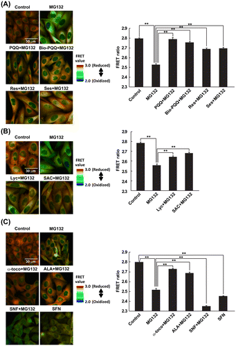
Fig. 2. Detection of mitochondrial ROS.
Notes: Mitochondrial ROS were detected using MitoSOX Red probe after 8-h treatment. Representative fluorescence images of MitoSOX Red are shown in the left panels. All images are the same magnification; scale bar = 50 μm. The quantitated relative fluorescence intensities are shown as the mean ± SEM of three independent measurements in the right panels. (A) Treatment with DMSO (control), MG132, PQQ + MG132, Bio-PQQ™ + MG132, Res + MG132 and Ses + MG132. (B) Treatment with Lyc + MG132 and SAC + MG132. (C) Treatment with α-toco + MG132, ALA + MG132, SFN + MG132 and SFN alone. ** p < 0.01.
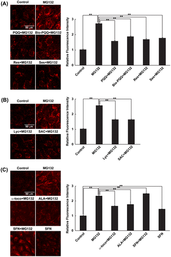
Fig. 3. Effect of dietary antioxidants against ROS generated in mitochondria.
Notes: Mitochondrial ROS was detected using MitoSOX Red probe. Cells were treated with respective antioxidants only after 4 h of MG132 treatment with total incubation time of 8 h unlike simultaneous treatment in Fig. . Reprehensive fluorescence images of MitoSOX Red is shown in upper panels. All images are the same magnification; scale bar = 50 μm. The quantitated relative fluorescence intensity is shown as the mean ± SEM of three independent measurements in lower panel. ** p < 0.01.
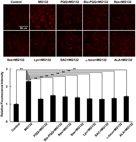
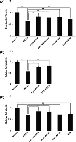
Fig. 4. MTT Cell viability assay.
Notes: Relative cell viability was determined by the MTT assay. Data are represented as relative fluorescence with respect to DMSO-treated cells. (A) treatment with DMSO (control), MG132, PQQ + MG132, Bio-PQQ™ + MG132, Res + MG132 and Ses + MG132. (B) treatment with Lyc + MG132, and SAC + MG132. (C) treatment with α-toco + MG132, ALA + MG132, SFN + MG132 and SFN alone. All of the treatments were carried out for 8 h. Quantitated values are shown as the mean ± SEM of three independent measurements. **: p < 0.01, NS = not significant.
Supplemental material