Figures & data
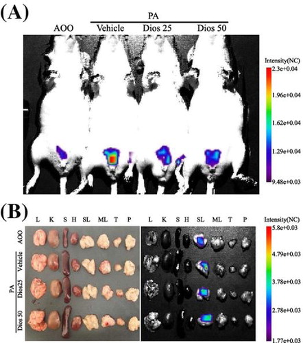
Fig. 1. Chemical structure of DG and ear morphological analysis of IL-4/Luc/CNS-1 Tg mice.
Notes: (A) DG consists of nonpolar aglycones attached via an ether bond to one or more monosaccharide moieties. (B) and (C) Ear thickness of mice in four groups was measured using a thickness gage and phenotypes were observed as described in the Materials and Methods. DG was administered via oral gavage for four weeks, while PA solution was repeatedly applied to the dorsum of the ear of IL-4/Luc/CNS-1 Tg mice during the same period. The vein in the ear was indicated by arrows. (D) After collection of blood serum from the abdominal vein of the mice, the IgE concentration was quantified by an enzyme-linked immunosorbent assay (ELISA). Data shown are the means ± SD (n = 5). a, p < 0.05 compared to the AOO treated group. b, p < 0.05 compared to the PA + Vehicle treated group. c, p < 0.05 compared to the PA + DG25 treated group.
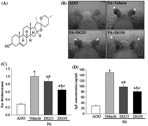
Fig. 2. Measurement of luciferase signal in the whole body (A) and each organ (B) of IL-4/Luc/CNS-1 Tg mice.
Notes: After cotreatment with PA and DG for 4 weeks, the luciferase signal was measured in AOO, PA + Vehicle, PA + DG25 and PA + DG50 treated mice using the Living Image software. The color overlay on the image represents the photons emitted per second from the organs in accordance with the pseudocolor scale shown next to the image. In this image, red indicates the highest number of photons per second, while blue indicates the lowest. L, lung; K, kidney; S, spleen; H, heart; SL, submandibular lymph node; ML, mesenteric lymph node; T, thymus; P, pancreas. (C) The level of luciferase signal emitted from whole body and SL were quantified using the Living Image software (Xenogen). Data shown are the means ± SD (n = 5). a, p < 0.05 compared to the AOO treated group. b, p < 0.05 compared to the PA + Vehicle treated group.
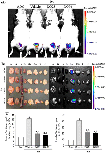
Fig. 3. Body and organ weight analysis.
Notes: (A) The body weight of mice in each subset group was measured with a chemical balance at the indicated time points. (B-D) Following final treatment, mice from each group were sacrificed under anesthesia, after which the spleen, thymus, and lymph nodes were harvested using a micro scissor and weighed on a chemical balance. Data shown are the means ± SD (n = 5). a, p < 0.05 compared to the AOO treated group. b, p < 0.05 compared to the PA + Vehicle treated group. c, p < 0.05 compared to the PA + DG25 treated group.
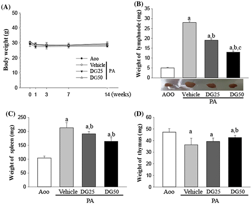
Fig. 4. Histopathological analysis of ear tissue.
Notes: Ear skin was collected from AOO, PA + Vehicle, PA + DG25, and PA + DG50 treated IL-4/Luc/CNS-1 Tg mice after repeated application of PA solution and administration of DG. The histopathological changes in the slide sections of ear tissue were identified by staining with hematoxylin and eosin (left and middle column) or toluidine blue (right column) followed by observation at 100× and 400× magnification. The infiltrated mast cells in the dermis of the ear tissue were indicated by arrow heads in the toluidine blue stained image. Data shown are the means ± SD (n = 5). a, p < 0.05 compared to the AOO treated group. b, p < 0.05 compared to the PA + Vehicle treated group. ND, not detected.
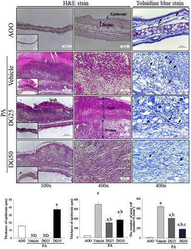
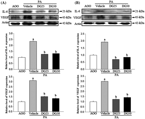
Fig. 5. Analysis of cytokine expression in ear tissues (A) and lymph nodes (B).
Notes: Western blot analysis of total protein was extracted from the lymph nodes and ear tissue of IL-4/Luc/CNS-1 Tg mice from subset groups. The levels of IL-6 and VEGF were detected with specific antibodies. The actin level is also shown as an endogenous control. The band intensity of IL-6 and VEGF protein was determined using an imaging densitometer and the relative level of each protein was calculated based on the intensity of actin protein as an endogenous control. Data represent the means ± SD from three replicates. a, p < 0.05 compared to the AOO treated group. b, p < 0.05 compared to the PA + Vehicle treated group.