Figures & data
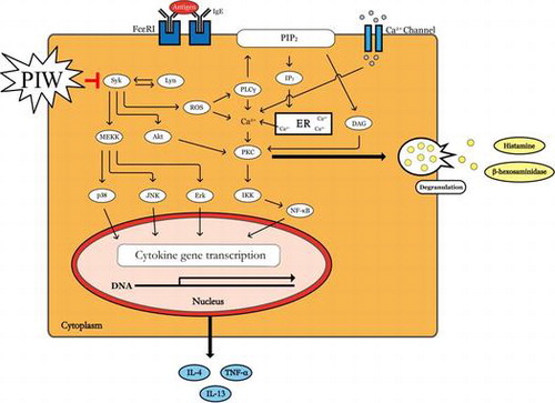
Fig. 1. Effects of PIW on IgE/Ag-induced RBL-2H3 cell viability and degranulation.
Notes: (A) Cytotoxicity. RBL-2H3 cells were incubated overnight in 96-well plates and treated with PIW at various concentrations (10, 25, 50, and 100 μg/mL) for 24 h. Cell viability was determined with the Ez-Cytox kit by measuring the absorbance at 450 nm. (B) Beta hexosaminidase release. RBL-2H3 cells were incubated overnight in 24-well plates with 400 ng/mL DNP-specific IgE. The medium was replaced with PIPES buffer containing the indicated concentration of PIW, and the cells were challenged with 50 ng/mL DNP-BSA. After 15 min, β-hexosaminidase release was determined from the absorbance measured at 405 nm. Data are expressed as mean ± SD (n = 3). **p < 0.01.
Fig. 2. Effect of PIW on IgE/Ag-induced passive cutaneous anaphylaxis.
Notes: Seven-week-old ICR mice were injected intradermally with DNP-IgE (0.5 μg) into the ear; after 24 h, ICR mice were intravenously injected with 200 μg DNP-BSA containing 3% Evans blue. PIW (50 or 100 mg/kg) or DPH (50 mg/kg) was orally administered 1 h before DNP-BSA, as indicated. (A) Representative photographs of the ears. (B) The dye was extracted overnight in 500 μL of formamide at 63 °C, and light absorbance was measured at 620 nm. Data are expressed as mean ± SD (n = 6). **p < 0.01.
Fig. 3. Effect of PIW on IgE/Ag-induced production of intracellular ROS.
Notes: (A) Scavenging of PIW according to DPPH assay. Cells were treated with PIW (10, 25, 50, 100 μg/mL), ethanol, and DPPH solution in 96-well plates and then incubated for 30 min without light. Optical density was determined at 517 nm. (B) Measurement of intracellular ROS. RBL-2H3 cells were incubated overnight in 6-well plates with 400 ng/mL DNP-specific IgE. The medium was replaced with the indicated concentration of PIW, and the cells were incubated with 10 μM DCF-DA at 37 °C for 30 min. The cells were then challenged with 50 ng/mL DNP-BSA. Representative images from a fluorescence microscope are shown. Data are expressed as mean ± SD (n = 3).
Fig. 4. Effect of PIW on IgE/Ag-induced phosphorylation of Syk, PLC-γ, Akt, Erk, and JNK.
Notes: RBL-2H3 cells were incubated and sensitized with 400 ng/mL DNP-specific IgE for 12 h and then treated with PIW at the indicated concentration. (A) Western blot analysis. Representative data of Western blot analyses for PLC-γ, Akt, Erk, and JNK. (B) Densitometric analysis of the expression of phosphorylated proteins. Data are expressed as mean ± SD (n = 3). *p < 0.05, **p < 0.01, and ***p < 0.001.
Fig. 5. Effect of PIW on IgE/Ag-induced expression of IL-4 and IL-13 mRNA.
Notes: RBL-2H3 cells were incubated and sensitized with 400 ng/mL DNP-specific IgE for 12 h and then treated with PIW for 30 min at the indicated concentration. DNP-BSA (50 ng/mL) was applied to IgE-sensitized RBL-2H3 cells for 1 h. Total RNA was isolated, and real-time PCR analysis was performed to detect IL-4 and IL-13 mRNA expression. Data are expressed as mean ± SD (n = 3). **p < 0.01.
Fig. 6. Effect of PIW on DNCB-induced AD-like ICR mice.
Notes: During induction of atopic dermatitis, 10 mg/mL PIW was applied to the dorsal skin of mice a total of 6 times over the course of 2 weeks. (A) Effect of PIW on DNCB-induced AD-like skin lesions in ICR mice. After inducing AD, a 10 mg/mL PIW solution (dissolved in a 3:1 mixture of acetone and olive oil) was applied to the dorsal skin of mice, a total of 6 times over a 2-week period. The mice were then sacrificed, and the dorsal hair of each mouse was shaved. (B) Skin severity score. Skin severity was assessed macroscopically in a blinded fashion. The development of AD-like skin dermatitis was scored as 0 (none), 1 (mild), 2 (moderate), or 3 (severe). The sum of the individual scores was defined as the dermatitis score. (C) Scratching frequency. The scratching behavior of each mouse was videotaped for 60 min every 2 days after sensitization. Biting of the dorsal skin and scratching with hind paws were observed during playback. (D) Histological analysis. Dorsal skin of AD-like ICR mice was isolated, sectioned, and stained with hematoxylin and eosin. Histological analysis was performed by light microscopy at 40× and 100× magnification. (E) IgE level in serum. Serum was collected immediately before sacrifice, and IgE levels were measured by sandwich ELISA using a mouse IgE kit according to the manufacturer’s instructions. Data are expressed as mean ± SD (n = 6). **p < 0.01.
Fig. 7. Effect of PIW on DNCB-induced cytokine levels in skin lesions in AD-like ICR mice.
Notes: Total RNA was extracted from dorsal skin tissue, and mRNA levels of TNF-α, IL-4, and IL-13 were determined by real-time PCR. Data are expressed as mean ± SD (n = 6). **p < 0.01.
Fig. 8. PIW suppresses IgE/Ag-stimulated degranulation by blocking the phosphorylation of Syk and associated downstream signal molecules.