1,607
Views
21
CrossRef citations to date
0
Altmetric
Analytical Chemistry

In vitro selection of DNA aptamers binding pesticide fluoroacetamide

, , , , , & show all
Pages 823-832 | Received 25 Sep 2015, Accepted 20 Nov 2015, Published online: 12 Feb 2016

Figures & data

Fig. 1. Structure formula of fluoroacetamide and its analog, N-fluoroacetyl glycine (A) and synthetic steps of N-fluoroacetyl glycine (B).
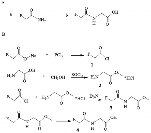
Fig. 1. Structure formula of fluoroacetamide and its analog, N-fluoroacetyl glycine (A) and synthetic steps of N-fluoroacetyl glycine (B).

Table 1. Sequences of the 40nt core regions of the cloned aptamers.

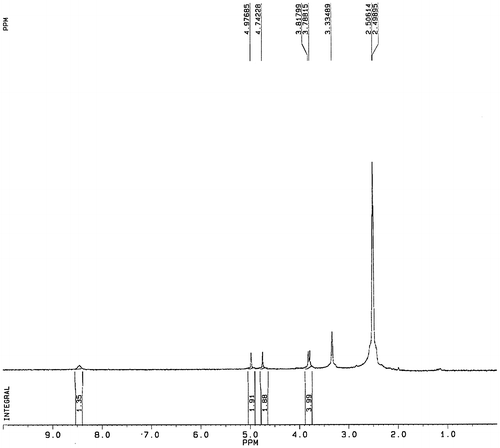
Fig. 2. 1H NMR spectra confirming the structure of fluoroacetamide analog.

Fig. 2. 1H NMR spectra confirming the structure of fluoroacetamide analog.

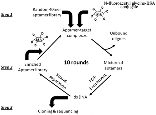
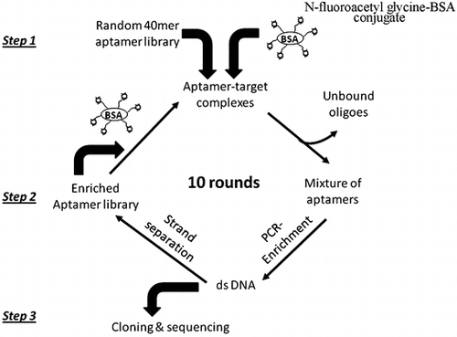
Fig. 3. Schematic representation of aptamer selection procedure.

Fig. 3. Schematic representation of aptamer selection procedure.

Fig. 4. Analysis of PCR products from each round of SELEX enrichment. PCR products were sampled after each round of enrichment and run on a 2% agarose gel.

Notes: M: molecular ladder (50–500 bp), Lanes 1-10: PCR products from enrichment rounds 1-10, and lane 11: no template control.
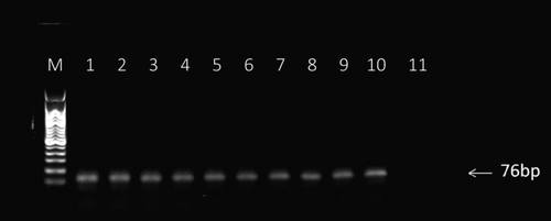
Fig. 4. Analysis of PCR products from each round of SELEX enrichment. PCR products were sampled after each round of enrichment and run on a 2% agarose gel.Notes: M: molecular ladder (50–500 bp), Lanes 1-10: PCR products from enrichment rounds 1-10, and lane 11: no template control.

Fig. 5. Affinity of the selected ssDNA to the target in each selection round. The initial ssDNA library was used as reference.
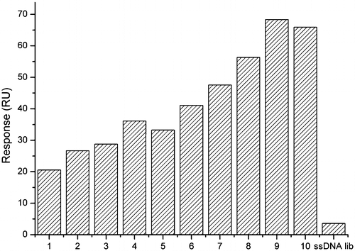
Fig. 5. Affinity of the selected ssDNA to the target in each selection round. The initial ssDNA library was used as reference.

Fig. 6. Multiple sequence alignments of the selected aptamers (without primers).
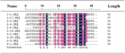
Notes: All positions with 75% or higher homology were highlighted and represented by lowercase letters in the consensus line. Identity = 66.89%. Numbers on the right-hand side indicate the length of each aptamer sequence. The number of nucleotides in the randomized region may have been changed by PCR mutations.
Fig. 6. Multiple sequence alignments of the selected aptamers (without primers).

Fig. 7. Predicted secondary structures of aptamer 1-1, 6-1, 2-8, and 1-17.
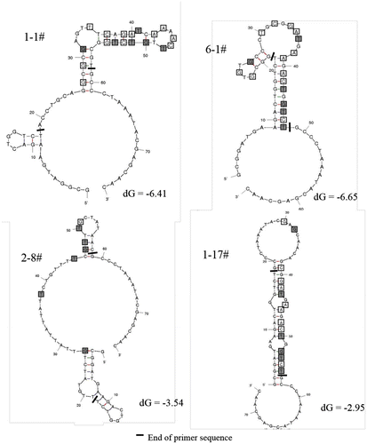
Note: The bases with high similarity were marked by square.
Fig. 7. Predicted secondary structures of aptamer 1-1, 6-1, 2-8, and 1-17.

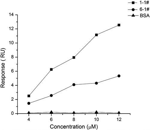
Fig. 8. Binding curves of aptamer 1-1 and 6-1 to N-fluoroacetyl glycine examined by SPR.

Fig. 8. Binding curves of aptamer 1-1 and 6-1 to N-fluoroacetyl glycine examined by SPR.

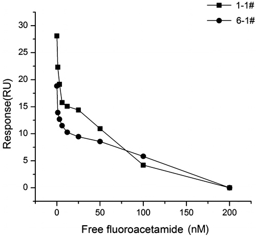
Fig. 9. Binding curves of aptamer 1-1 and 6-1 in the SPR-based inhibition assay.

Fig. 9. Binding curves of aptamer 1-1 and 6-1 in the SPR-based inhibition assay.

Fig. 10. Characterization of specificity of aptamer 1-1 for fluoroacetamide. The binding affinity of aptamer 1-1 to fluoroacetamide and other structurally similar molecules was tested by a dipstick assay based on lateral-flow technology. The fluorescence intensity at test line for each target was measured. The buffer solution without any target was used as blank.
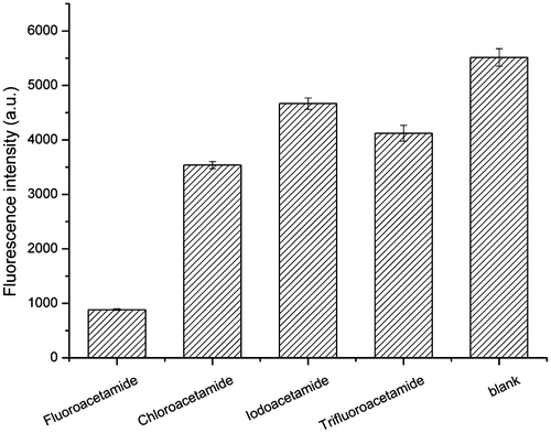
Fig. 10. Characterization of specificity of aptamer 1-1 for fluoroacetamide. The binding affinity of aptamer 1-1 to fluoroacetamide and other structurally similar molecules was tested by a dipstick assay based on lateral-flow technology. The fluorescence intensity at test line for each target was measured. The buffer solution without any target was used as blank.
Reprints and Corporate Permissions

Please note: Selecting permissions does not provide access to the full text of the article, please see our help page How do I view content?

To request a reprint or corporate permissions for this article, please click on the relevant link below:

Academic Permissions

Please note: Selecting permissions does not provide access to the full text of the article, please see our help page How do I view content?

Obtain permissions instantly via Rightslink by clicking on the button below:

If you are unable to obtain permissions via Rightslink, please complete and submit this Permissions form. For more information, please visit our Permissions help page.