
Figures & data
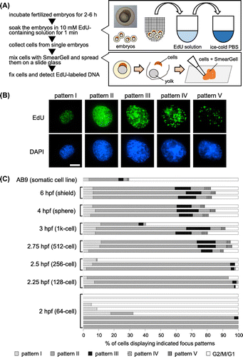
Fig. 1. Visualization of replication dynamics in Zebrafish embryo.
Notes: (A) Overview of experimental procedure. Zebrafish embryos were labeled with EdU, and subsequently, replication sites were visualized in single embryo-derived cells through fluorescence-mediated detection. (B) Spatial distribution patterns of DNA replication sites (patterns I-V) in zebrafish cell nuclei collected from a 6 hpf embryo. EdU-labeled DNA was detected by a click reaction with the green fluorescent Alexa Fluor 488 azide (above), and nuclear DNA was stained with DAPI (below). Scale bar, 5 μm. (C) Frequency of replication focus patterns at each developmental stage. Cells were isolated from EdU-labeled single embryos at each developmental stage, and replication sites were visualized as described in (B). The percentage of cells displaying each focus pattern (I-V) and unlabeled cells were scored (2 hpf: n = 18–56 cells/embryo, average 34; 2.25 hpf: n>100/embryo, 2.5–6 hpf: n>200/embryo). Each horizontal bar represents the result from a single embryo.
Supplemental material