Figures & data
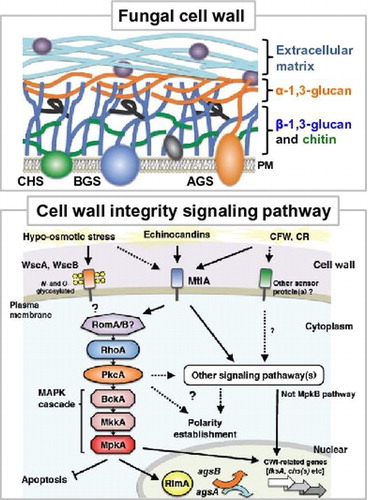
Fig. 1. Schematic illustration of cell wall architecture in Aspergillus species.
Notes: The cell wall central core is mainly composed of β-1,6-branched β-1,3-glucan crosslinked to chitin, and amorphous α-1,3-glucan is present in the cell wall outer layer. Polysaccharides, such as galactosaminogalactan and galactomannan, and proteins, such as GPI-anchored and surface proteins, are also present in cell wall. Abbreviations: AGS, α-1,3-glucan synthase; BGS, β-1,3-glucan synthase; CHS, chitin synthase; and ECM, extra cellular matrix.

Fig. 2. Chemical structure of polysaccharides associated with fungal cell wall.
Note: (A) linear α-1,3-glucan (B) β-1,6-branched β-1,3-glucan (C) Chitin.
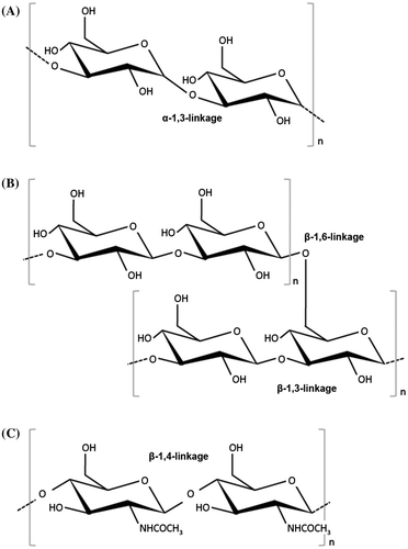
Fig. 3. Phylogenetic tree of α-1,3-glucan synthases (A), β-1,3-glucan synthases (B), and chitin synthases (C) in yeast and filamentous fungi.
Notes: The tree was constructed using the neighbor-joining method based on alignment of amino acid sequences. An, Aspergillus nidulans; Ao, A. oryzae; Af, A. fumigatus; Anig, A. niger; Nc, Neurospora crassa; Mg, Magnaporthe grisea; Bc, Botrytis cinerea; Ss, Sclerotinia sclerotiorum; Pr, Penicillium rubens; Ca, Candida albicans; Sp, Schizosaccharomyces pombe; and Sc, Saccharomyces cerevisiae.
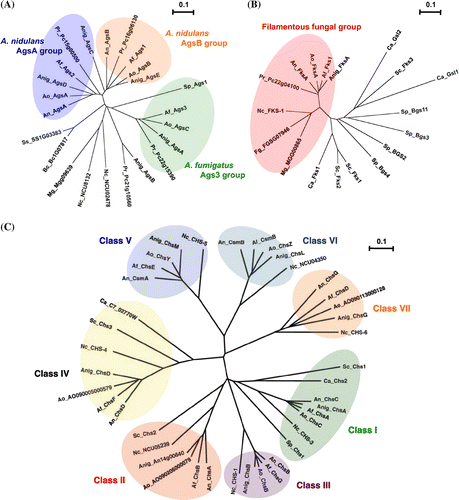
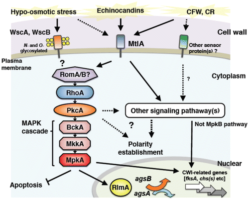
Fig. 4. Schematic model of cell wall stress signaling in A. nidulans.
Notes: Based on study results, we hypothesize that A. nidulans has the following CWI signaling system: (1) Putative sensor proteins in the CWI signaling pathway, WscA, WscB, and MtlA, play an important role in CWI signaling under hypo-osmotic conditions, but WscA and WscB are not essential for MpkA-RlmA signaling. (2) PkcA is involved in the CWI pathway in A. nidulans. In addition, PkcA plays a role in suppression of apoptosis induction via the MpkA pathway, but not in polarity establishment, during hyphal growth independent of the MpkA pathway under heat-stress conditions. (3) Expression of agsA and agsB is dependent on MpkA and partly dependent on RlmA. (4) Other CWI-related genes, such as fksA, gelA, gelB, chsA, chsB, chsC, chsD, csmA, and csmB, are independent or partly dependent of the MpkA-RlmA system. The CWI pathway mainly regulates transcription of α-1,3-glucan biogenesis-related genes. Transcripts of β-1,3-glucan and chitin biogenesis-related genes are mainly regulated by other unknown signals that might be activated by a cell wall stress, such as echinocandin (micafungin) treatment.