
Figures & data

Table 1. The Neurospora crassa strains used in this study.
Fig. 1. Comparison of the growth and morphology of mak-1, mak-2, and os-2 disruptants of Neurospora crassa.
Notes: (A) Hyphal morphology of the wild-type, mak-1, mak-2, and os-2 disruptants on Vogel’s medium N (VN). Photographs were taken when the mycelia reached the edge of the plate. (B) Relative growth rates of the wild-type, mak-1, mak-2, and os-2 disruptants. Each strain was grown on VN for 24 h at 28 °C and the length of the mycelia of the wild-type was set as 100%. (C) Sensitivity of mak-1, mak-2, and os-2 disruptants to micafungin, a beta-1,3-glucan synthase inhibitor. Conidia (10-fold dilution series) of each strain were inoculated on Vogel’s minimal medium plates containing 1% sorbose, 0.2% sucrose, and 1 μg/ml micafungin for 48 h at 28 °C.
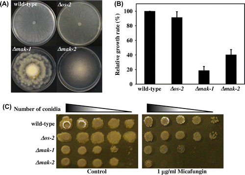
Fig. 2. Phosphorylation of MAK-1 and MAK-2 MAPKs is induced by micafungin.
Notes: (A) Phosphorylation status of MAK-1 and MAK-2 from conidial germination to hyphal development in the wild-type strain. (B) Phosphorylation status of MAK-1 and MAK-2. Mycelia were cultivated in liquid Vogel’s medium N for 16 h at 28 °C, and then exposed to 1 μg/ml micafungin for 2 h. −, untreated control; +, micafungin-treated. In these experiments, an equal amount of total cell protein (50 μg per lane) were separated by 10% SDS–PAGE and subjected to Western blotting analysis with the following antibodies: anti-ERK1/2 for total MAK-2, anti-phospho-p44/42 MAPK for phosphorylated MAK-1 and MAK-2.
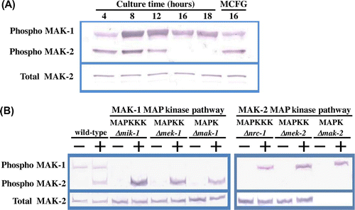
Table 2. Expression analysis of 12 cell wall biogenesis-related genes by quantitative real-time RT-PCR.
Table 3. Microarray analysis of genes upregulated by micafungin treatment in wild-type strain.
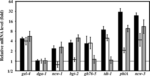
Fig. 3. Relative mRNA levels of selected genes in response to micafungin.
Notes: Relative quantities of mRNA of gel-4, dga-1, ncw-1, bgt-2, gh76-5, tdt-1, phiA, and ncw-3 in response to micafungin (1 μg/ml for 2 h) in the wild-type (black boxes), Δmak-1 (white boxes), and Δmak-2 (gray boxes). Quantification of mRNA by real-time RT-PCR is presented as a relative value for each treatment by comparison with the mRNA levels in the untreated control. We had already reported that gel-4 was significantly upregulated by micafungin treatment in the wild-type, Δmak-1, and Δmak-2 strains.Citation26) Three independent experiments were carried out. Standard deviations are indicated as error bars.
Supplemental material