Figures & data
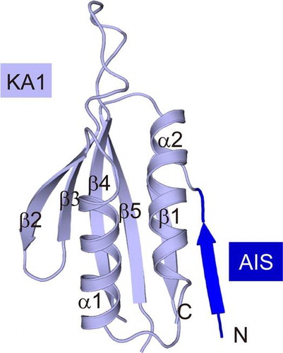
Fig. 1. Biochemical characteristics of SAD-B KD-UBA.
Notes: (A) Schematic diagram of mouse SAD-A/B. The structural elements of SAD-B and SAD-A are, respectively, colored and indicated. The sequence of SAD-B AIS is provided, with the key residues highlighted in red. (B) Time course of phosphorylation/activation of SAD-B KD-UBA by LKB1. top: Phosphorylation of SAD-B Thr189 by LKB1 analyzed using an anti-AMPK-pT172 antibody; bottom: Plots of initial rate of SAD-B catalyzed Cdc25C phosphorylation versus the phosphorylation time. At indicated time intervals, 10 nM indicated SAD-B KD-UBA was added in the reaction buffer containing 20 μM Cdc25C peptide. (C) Time course of SAD-B KD-UBA catalyzed phosphorylation of Cdc25C. Reactions were initiated by adding 10 nM indicated SAD-B KD-UBA to the reaction buffer with 20 μM Cdc25C peptide. (D) Plots of initial velocity of SAD-B-catalyzed Cdc25C phosphorylation versus the Cdc25C concentration. The solid lines represent the best fitting results to the Michaelis–Menten equation. 5 nM phosphorylated SAD-B KD-UBA was added in the reaction buffer with different concentration of Cdc25C peptide.
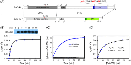
Table 1. Data collection and refinement statistics.
Fig. 2. Overall structure of AIS-KA1 of mouse SAD-B.
Notes: (A) Overall structure of molecule A. The color scheme follows that in Fig. (A). (B) Cartoon representation of AIS-KA1. The structural elements of molecule A and molecule B are, respectively, colored as follows: AIS (dark blue and orange, respectively) and KA1 (light blue and gray yellow, respectively) (C) Oligomerization state of AIS-KA1 from SAD-B at different concentrations by gel filtration chromatograph. AIS-KA1 was eluted with retention volume corresponding to an apparent molecular weight of ~15 kDa. (D) Comparison of the KA1 structures of molecules A and B in the crystallographic SAD-B dimer and that from the SAD-A complex of KD-UBA and AIS-KA1 (PDB ID: 4YOM).
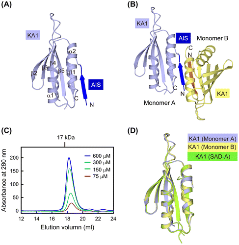
Fig. 3. Analysis of the AIS sequence in the free AIS-KA1 structure.
Notes: (A) Sequence alignment of AIS-KA1 fragments from SAD kinases generated by Clustal W. Residues forming the AIS are boxed in dark blue, and the key inhibitory residues in AIS are indicated by asterisks. Residues forming the hydrophobic core of KA1 are boxed in black. Secondary-structure elements are labeled. (B) Superposition of the two molecules within the asymmetric units of SAD-B AIS-KA1. (C) The Fo-Fc omit map (contoured at 1.5 σ) shows electron density for the C-terminal part of AIS (597GNFISLD603). (D) Close-up view of the compared AIS of molecule A and B. The residues 597–603 are shown as sticks, and residue Phe599 and Ile600 are labeled.
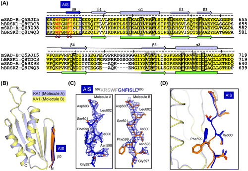
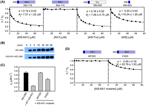
Fig. 4. The conserved AIS inhibits the kinase activity of SAD-B.
Notes: (A) Trans-inhibition of different C-terminal fragments on the activity of the pKD-UBA fragment (10 nM). The continuous curve was the best-fit to the non-competitive model using equation vi /v0 = (Ki + a[I]/(Ki + [I]), where Ki and a are the apparent inhibition constant and residual activity, respectively. (B) Time course of LKB1-catalyzed phosphorylation of KD-UBA in isolation and in complex with AIS-KA1. (C) Effects of the AIS mutations on the trans-inhibition of AIS-KA1 on the pKD-UBA activity. The assays were performed with 10 nM pKD-UBA and 50 μM AIS-KA1 mutants (mean ± SEM, n = 3). (D) Trans-inhibition of AIS-KA1 mutants (W595D/F596D and F599D/I600D) on the activity of the pKD-UBA fragment.