Figures & data
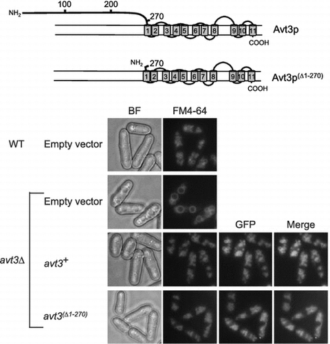
Fig. 1. Topology models of Avt3p and immunoblot analysis of GFP–Avt3p and GFP–Avt3p(Δ1–270) expressed in S. pombe avt3∆ cells.
Notes: (A) A topology model of S. pombe Avt3p predicted by the SOSUI program. (http://harrier.nagahama-i-bio.ac.jp/sosui/sosui_submit.html). Four phosphorylated residues in the N-terminal region were identified by the global analysis of phosphorylation, and the locations of these residues are indicated as [P]. A model of Avt3p with its N-terminal region deleted is also shown. (B) Left panel: protein samples were prepared from S. pombe avt3Δ cells expressing GFP–avt3+ or GFP–avt3(Δ1–270) and then analyzed by immunoblotting using anti-GFP serum. Asterisks indicate putative degradation products. Right panel: Coomassie Brilliant Blue-stained gel is shown as loading control.
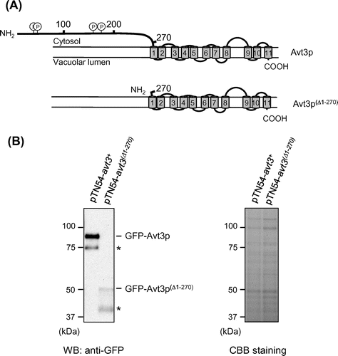
Fig. 2. Effect of deletion of the N-terminal region of Avt3p on its subcellular localization and vacuolar morphology in S. pombe cells.
Notes: S. pombe avt3∆ cells expressing GFP–avt3+ or GFP–avt3(Δ1–270) were grown in EMM medium without leucine and thiamine for 20 h, and vacuolar membranes were stained with FM4–64. BF, bright field; Bar, 5 μm.
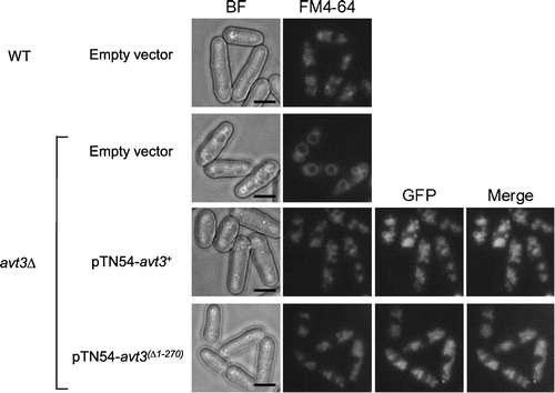
Fig. 3. Effect of deletion of the N-terminal region of Avt3p on vacuolar amino acid composition in S. pombe cells.
Notes: Vacuolar pools of S. pombe were prepared and analyzed with an amino acid analyzer as described in “Materials and methods.” Results are mean ± SD from three independent experiments. Statistically significant differences between groups were determined by two-tailed Student’s t-test. *, p < 0.05: S. pombe wild-type cells carrying empty plasmid (white bar), avt3∆ cells carrying empty plasmid (black bar), pTN54–avt3+ (light gray bar), or pTN54–avt3(Δ1–270) (dark gray bar).
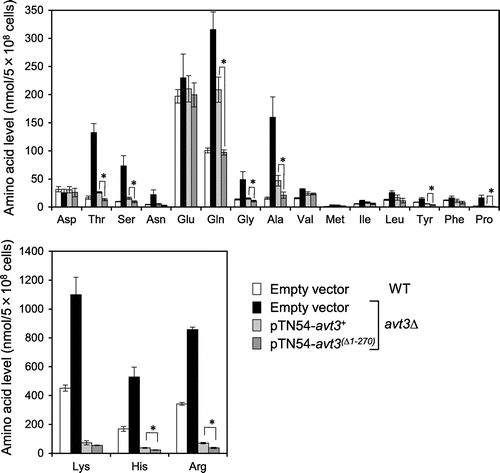
Fig. 4. Effect of deletion of the N-terminal region of Avt3p on spore formation of S. pombe.
Notes: (A) S. pombe KJ100-7B avt3∆ cells expressing GFP–avt3+ or GFP–avt3(Δ1–270) were grown in EMM medium without leucine and thiamine, and then grown on the MEA. After 24 h (Day1) or 48 h (Day2), cells were collected and then observed with fluorescent microscope. BF, bright field; Bar, 5 μm. (B) After culturing for 3 days in sporulation medium, the number of total cells and spores was counted under a microscope to calculate the sporulation efficiency. Results are means ± SD from three independent experiments. Statistically, significant differences between groups were determined by two-tailed Student’s t-test. *, p < 0.05.
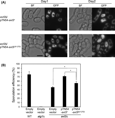
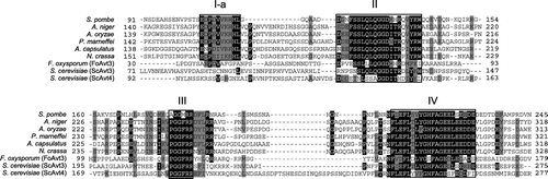
Fig. 5. Conserved sequence elements in the N-terminal hydrophilic regions of fungal Avt3/Avt4 homologs.
Notes: The amino acid sequence of N-terminal hydrophilic region of S. pombe Avt3p was aligned with those of the homologs of A. niger, A. oryzae, P. marneffei, A. capsulatus, N. crassa, F. oxysporum, and S. cerevisiae by CLUSTALW software. Identical and similar residues are represented as black and gray boxes, respectively. The clusters of highly conserved residues (I-a, II, III, and IV) in the Avt3/Avt4 homologs are shown.