Figures & data
Table 1. Primers used in this study.
Fig. 1. The expression profile of EWS during the early stages of neuronal differentiation.
Notes: (A) P19 cells were induced to differentiate with 5 μM all-trans-retinoic acid (RA). Real-time detection of PCR products was performed using a Light Cycler 480 Real-Time PCR System (Roche). Hypoxanthine guanine phosphoribosyl transferase was used as an internal control to normalize the data. Samples were quantified relative to the Ct (using the 2−ΔΔCT method,Citation19) where Ct is the threshold cycle value) of the internal control. Each experiment was repeated at least three times and statistical analysis was conducted using the Student’s t-test. Data are the mean ± standard deviation (*p < 0.01 vs. day 0). (B) P19 cells were induced to differentiate with 5 μM RA. Whole-cell lysates were prepared in 10 mM Tris-HCl (pH 7.4) containing 0.5% Nonidet P-40, 150 mM NaCl, and protease inhibitor cocktail (Nacalai Tesque), incubated for 5 min, and centrifuged at 13,200 rpm for 15 min. Proteins were separated in 10% SDS–PAGE. Representative western blot of whole-cell lysates probed with the indicated antibodies. Anti-EWS (G-5; Santa Cruz Biotechnology), anti–β3-tubulin (TU-20; Santa Cruz Biotechnology), and anti–α-tubulin (B-5-1-2; Sigma-Aldrich) were used. α-Tubulin was used as a loading control.
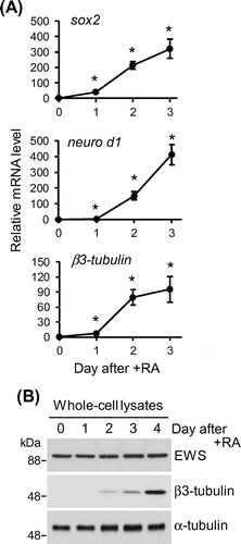
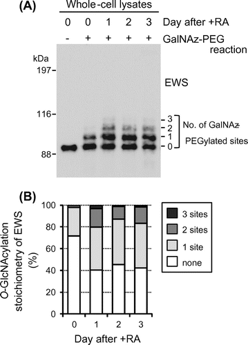
Fig. 2. O-GlcNAcylation stoichiometry of EWS is regulated by neuronal differentiation in P19 cells.
Notes: (A) Monitoring of O-GlcNAcylated EWS species during neural differentiation. P19 cells were induced to differentiate with 5 μM RA. Whole-cell lysates were prepared and reacted chemoenzymatically with polyethylene glycol (PEG) mass tags to label O-GlcNAcylated proteins. Briefly, azide-modified galactose (GalNAz) was enzymatically transferred to terminal GlcNAc residues by a mutant β-1,4-galactosyltransferase (GalTY289L) using a Click-iT O-GlcNAc Enzymatic Labeling System (Thermo Fisher Scientific). The PEG mass tag was then reacted with GalNAz by click chemistry using a Click-iT Protein Analysis Detection kit (Thermo Fisher Scientific) according to the manufacturer’s protocol, with the exception that the alkyne-labeled biotin was replaced with 5 kDa mPEG-Alkyne. GalNAz-PEG-labeled proteins were resolved by SDS–PAGE and immunoblotted with anti-EWS antibody (G-5). As a negative control, reactions were performed in the absence of GalTY289L (-GalNAz-PEG reaction). The number on the right side of the blot panel indicates the GalNAz-PEG-modified site number of the corresponding band. (B) Quantification of the O-GlcNAcylation stoichiometry of EWS. The stoichiometry of O-GlcNAc glycosylation of EWS was determined by densitometric analysis of the relative intensities of the bands in (A).