Figures & data
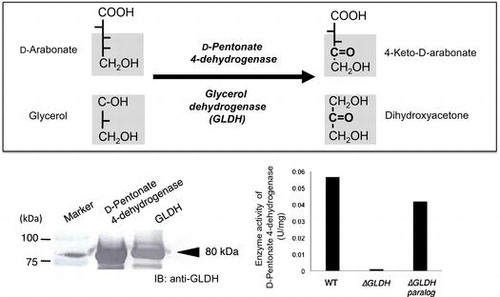
Fig. 1. Effects of EDTA on D-pentonate 4-dehydrogenase.
Notes: D-arabonate and D-ribonate were incubated with native (N), EDTA-treated (E), and the reactivated membrane with PQQ and Ca2+ (R). The reaction mixture contained 50 μL of membrane solution of 20 mg of protein/mL in 10 mM acetate buffer pH 5.0, and 50 μL of 0.5 M substrate. The enzyme reaction was carried out for 18 h at 30˚C with gentle shaking. To the reaction mixture, trichloroacetic acid was added to 10% (v/v). The reaction mixtures were spun down by a tabletop centrifuge. An aliquot of the supernatant was spotted on a TLC plate and developed with a solvent system of n-butanol : formic acid : water = 4 : 1 : 1.5. Detection of reaction products possessing ketones was done by spraying an alkaline ethanol solution of 2,3,5-triphenyltetrazolium chloride over the TLC plate.
Fig. 2. Effects of EDTA on D-fructose 5-dehydrogenase.
Notes: The native membrane and EDTA-treated membrane were incubated with D-fructose by adding either PQQ or Ca2+, and both of PQQ and Ca2+. D-fructose (F) and 5-keto-D-fructose (KF) were used as the authentic standard. Analytical conditions are the same as in Fig. .
Fig. 3. CM-Toyopearl chromatography of D-pentonate 4-dehydrogenase.
Notes: After the solubilized enzyme solution was dialyzed against 5 mM acetate buffer, pH 4.0, containing 5 mM CaCl2 and 0.2% Mydol 10, the enzyme solution was applied to a CM-Toyopearl column (1.5 x 15 cm), which was equilibrated with the same acetate buffer. After washing the column with the buffer, the enzyme was eluted by a linear gradient made by 750 ml each of 10 mM to 35 mM CaCl2. D-Pentonate 4-dehydrogenase was assayed with PMS-DCIP as the electron acceptors. Enzyme activities of 4-aldopentose 4-dehydrogenase, aldehyde dehydrogenase (ALDH), and alcohol dehydrogenase (ADH) were assayed with K-ferricyanide as the electron acceptor.Citation12)
Fig. 4. Gel filtration, absorption spectrum (A) and SDS-PAGE (B) of purified D-pentonate 4-dehydrogenase.
Notes: (A) After the pooled enzyme fraction was concentrated with sucrose, a part of the enzyme solution was applied to a Sephadex G-200 column (1.5 × 120 cm), which had been equilibrated with 5 mM acetate buffer, pH 4.0, containing 5 mM CaCl2 and 0.2% Mydol 10, and fractionated by every 3 mL fraction. Absorption spectrum of purified D-pentonate 4-dehydrogenase is shown in the panel. Purified enzyme solution (1.7 mg/mL) in acetate buffer, pH 4.0, was used. Absorption spectrum was taken by a Hitachi Model 200-10 spectrophotometer. (B) SDS-PAGE of purified D-pentonate 4-dehydrogenase (5 μg) was developed. The dual color standard proteins of molecular masses from 250 kDa to 15 kDa (Bio-Rad, Hercules, CA, U.S.A.) were used as references for measurement of the molecular masse of the enzyme.
Fig. 5. Reaction versatility of GLDH to D-pentonate, D-fructose, D-psicose, and D-erythronate on the basis of the Bertrand–Hudson’s rule.
Notes: GLDH has been known to oxidize various compounds, such as glycerol, meso-erythritol, D-arabitol, D-sorbitol, D-mannitol, and D-gluconate to dihydroxyacetone, L-erythrulose, D-xylulose, L-sorbose, D-frucose, and 5-keto-D-gluconate according to Bertrand–Hudson’s rule.Citation15) The present study revealed that D-pentonate, D-fructose, D-psicose, and D-erythronate are also oxidized to 4-keto-D-pentonate, 5-keto-D-fructose,Citation25) 5-keto-D-psicose, and 3-keto-D-erythronate by GLDH reaction. Asterisks indicate the putative structure because the authentic standards are not available.
Fig. 6. Immunoblotting analysis of D-pentonate 4-dehydrogenase and glycerol dehydrogenase (GLDH).
Notes: The purified enzyme (5 μg) was developed on SDS-PAGE and then transferred to a PVDF membrane. The enzyme band was visualized using anti-GLDH antibody prepared with purified GLDH from G. thailandicus NBRC 3257.
Fig. 7. Effect of gene disruption on D-pentonate 4-dehydrogenase activity.
Notes: The membrane fraction of G. thailandicus NBRC 3255 and it variants were prepared by suspending with 5 mM Tris-HCl, pH 7.0. The enzyme activity for D-arabonate (final conc. 50 mM) was measured by K-ferricyanide reductase assay in 50 mM acetate buffer, pH 4.0, after reactivation with 5 mM CaCl2 and 5 µM PQQ.