
Figures & data
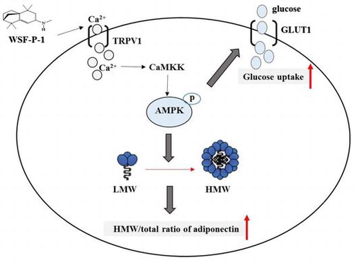
Fig. 1. WSF-P-1 activates AMPK by increasing intracellular calcium concentrations in 293T cells.
Notes: (A) Structure of WSF-P-1. (B) 293T cells were treated with WSF-P-1 in the presence or absence of 10 μM Compound C (CC) for 1 h. The cell lysates were subjected to SDS-PAGE and western blot analysis using antibodies specific for phospho-AMPK or total AMPK. The results are representative of three independent experiments with similar results. (C) The amount of pAMPK and AMPK was quantified using the NIH ImageJ software. The pAMPK/AMPK ratio (expressed as the mean ± SD, n = 3) was presented relative to the control (0 μM WSF-P-1 treated with or without CC). **, p < 0.01. (D) 293T cells were treated with WSF-P-1 or 40 μM evodiamine (Evo) for 1 h. After incubation with Fluo-3, the cells were analyzed on a flow cytometer using FL-1 as a detector. Relative intracellular calcium concentrations were calculated from the ratio of the geometric mean values of the FL-1 peak generated from WSF-P-1-treated cells against the control (0 μM WSF-P-1).
Fig. 2. WSF-P-1 activates AMPK via the Ca2+-dependent CaMKK signaling pathway.
Notes: (A) 3T3-L1 cells were pretreated with 10 μM Compound C (CC), 10 μM capsazepine (CPZ), or 1 μg/mL STO-609 (STO) for 1 h followed by treatment with WSF-P-1 for another hour. pAMPK and total AMPK were detected. (B) The AMPK/pAMPK ratio was calculated and presented relative to the control (0 μM WSF-P-1 treated with or without the same inhibitor). **p < 0.01.
Fig. 3. WSF-P-1 promotes adiponectin multimerization in 3T3-L1 adipocytes.
Notes: (A) 3T3-L1 adipocytes were treated with WSF-P-1 in the presence or absence of 10 μM capsazepine (CPZ) or 1 μg/mL STO-609 (STO) for 48 h. The cell lysates were subjected to 2–15% gradient gel electrophoresis under non-reducing and non-heat-denaturing conditions to detect the three oligomeric forms of adiponectin (LMW, MMW, and HMW) (top panel). The amount of total adiponectin (Total Ad), or actin was detected with antibodies against the N-terminal peptide of adiponectin, or β-actin, respectively. (B) The amounts of each oligomer and total adiponectin were quantified and the HMW/total ratio of adiponectin was calculated. The results were presented as the ratio of HMW/total relative to the control (0 μM WSF-P-1 treated with or without the same inhibitor). **p < 0.01. (C) 3T3-L1 adipocytes were transfected with AMPKα1 siRNA or control siRNA. Forty-eight hours after transfection, the cells were treated with WSF-P-1 for another 48 h. (D) The HMW/total ratio of adiponectin was calculated and presented as described in Fig. 3B.
Fig. 4. WSF-P-1 inhibits preadipocyte differentiation.
Notes: 3T3-L1 preadipocytes were induced to differentiate in the presence of WSF-P-1 or berberine (BBR) for 8 days. (A) The cells were stained with Oil Red O. Quantification of the staining results was presented in (B). Absorbance was detected at 490 nm. Results were presented relative to the control (0 μM WSF-P-1 or 0 μM BBR) *p < 0.05; **p < 0.01.
Fig. 5. WSF-P-1 promotes glucose uptake.
Notes: (A) 3T3-L1 adipocytes were treated with WSF-P-1 or berberine (BBR) in the presence or absence of 10 μM Compound C (CC) for 24 h. Glucose uptake was measured by a bioluminescent assay, as described in Materials and methods. The results were presented relative to the control (0 μM WSF-P-1 treated with or without CC). *p < 0.05; **p < 0.01. (B) Cells were treated with WSF-P-1 or BBR for 6 h. Total membrane proteins were isolated and analyzed by western blot using anti-GLUT1 antibody. The results were presented relative to the control (0 μM WSF-P-1).
Supplemental material